
Page 1194 - Hall et al (2015) Principles of Critical Care-McGraw-Hill
P. 1194
CHAPTER 88: Coma, Persistent Vegetative State, and Brain Death 831
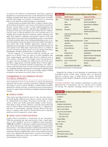
in injuries to the midbrain or medial thalami, which lead to significant TABLE 88-3 Useful Physical Examination Findings in Comatose Patients
impairment of consciousness but none or only minimal focal neurologic
findings. Traumatic brain injury with diffuse axonal injury can lead to Exam Focus Specific Features Suggested Condition
petechial hemorrhages and ischemia or inflammatory to necrotizing Skin Petechiae, splinter hemorrhage Coagulopathy; SBE
lesions of the midbrain, resulting in impaired consciousness.
Cerebral masses can produce either direct or indirect (uncal or tento- Icteric Hepatic encephalopathy
rial herniation) displacement and torsions of the midbrain and reduced Needle tracks Drug overdose or withdrawal
alertness. Sudden injury to either or both cerebral hemispheres may Cyanotic Hypoxemia
produce impaired consciousness, indicating that wakefulness has no
hemispheric dominance and requires some cerebral function. Bilateral, Lymph nodes Adenopathy Infectious etiologies;
immunocompromised hosts
extensive acute or subacute damage to the cortex and white matter, for
example, due to trauma, hypoxia, or infection, impairs activation of the Head Contusion; postauricular ecchymosis Trauma
upper RAS, leading to impaired consciousness. However, focal large (Battle sign)
cortical (lobar) areas can be injured and initially not affect consciousness VP shunt Hydrocephalus; shunt malfunction
at all until secondary injury from swelling and bleeding occurs, as evi- Eyes Periorbital ecchymosis (raccoon eyes) Trauma
denced by patients with penetrating wounds of the cerebral hemispheres
who remain fully awake. Similarly, degenerative disorders such as Papilledema Increased intracranial pressure
Alzheimer disease generally do not or only minimally affect the RAS and Ears Hemotympanum Trauma
these patients remain fully awake. Hypersomnia refers to a condition of Nose Excessive discharge Trauma
excessive drowsiness and sleep. It may occur in the setting of narcolepsy,
hypothalamic disorders, sleep disorders, or psychiatric illness. Neck Stiff Subarachnoid hemorrhage, infection
Several categories of consciousness-impairing mechanisms and Enlarged thyroid Dysthyroidism
lesions can be defined. First, one cause is an easily identifiable mass Cardiovascular Arrhythmia, etc Hypoxic/ischemic encephalopathy
lesion compressing the upper RAS either directly or indirectly (such as
tumor, abscess, meningitis, or hemorrhage); second, discrete lesions of Abdomen Small hard liver Hepatic encephalopathy
the upper brain stem (examples are outlined above) may be the cause; Miscellaneous Acetone, alcohol breath Ketoacidosis; alcohol intoxication
and third, a larger group of patients includes those in whom suppres- Fever Infection
sion of the RAS is induced by metabolic derangements, toxic states, or Tongue laceration; incontinence Postictal state
seizures. Such functional causes of coma may be reversible by correcting
the underlying metabolic derangement or removing the offending drug. SBE, subacute bacterial endocarditis; VP, ventriculoperitoneal.
“Metabolic” coma is likely the most common etiologic category resulting
in impaired consciousness in medical critical care units. Frequently the etiology of acute depression in consciousness in the
hospitalized patient includes sepsis, acid-base, fluid, and electrolyte
EXAMINATION OF THE COMATOSE PATIENT: disorders, or hepatic, renal, or cardiac failure or seizures. Therefore,
A CLINICAL APPROACH laboratory studies should be obtained to exclude metabolic and endo-
crine causes.
Acute depression in level of consciousness is a critical, life-threatening
emergency that requires a systematic approach for evaluation of etiology. Neurologic Examination: The neurologic examination in a patient with
The causes of coma are myriad. Therefore, a reliable history should be depressed level of consciousness can be a valuable tool to localize
obtained from family, witnesses, or medical personnel, and examination the etiology. The important neurologic features include (1) level of
should seek representative localizing neurological and general physical
findings. TABLE 88-4 Neurologic Assessment of the Comatose Patient
■ HISTORICAL FEATURES Level of consciousness
Clues can be ascertained from the onset of coma. An acute onset in a Arousability
previously healthy individual may indicate a cerebral vascular etiology Content
(ie, subarachnoid hemorrhage, intracerebral hemorrhage, or hemi-
spheric or brain stem stroke), generalized epileptic activity, traumatic Brain stem function
brain injury, or drug overdose. Likewise, a subacute deterioration may Respiratory rate and pattern
point to systemic illness, evolving intracranial mass, or a degenerative, Blood pressure and heart rate
infectious, or paraneoplastic neurologic disorder. Moreover, the dura- Pupil size and reactivity
tion of a comatose state should be documented because it may have
predictive value for prognosis in certain causes. Eye position and movements
■ GENERAL CLINICAL FEATURES AND PROTOCOL Doll’s eyes maneuver
Careful clinical examination is irreplaceable by any investigation. Cold caloric testing
Corneal reflexes
While the assessment may take some period of time, a quick assess-
ment of ABCs should be carried out in addition to basic resuscitation Facial symmetry
(Tables 88-3 and 88-4). A subsequent careful assessment should confirm Motor function
and evaluate the presence and extent of impairment of consciousness Posturing
and awareness. Core body temperature should be determined as hypo-
thermia can be seen in drug overdose, brain death, or acute spinal cord Tone
transection. Moreover, hyperthermia can be seen in infection; traumatic Spontaneous movements
brain injury; subarachnoid, intracerebral, or pontine hemorrhage; and Withdrawal to noxious stimulus
hypothalamic dysfunction. If there is no evidence of neck trauma, signs
of meningismus must be elicited. Deep tendon reflexes
section06.indd 831 1/23/2015 12:56:20 PM