
Page 1200 - Hall et al (2015) Principles of Critical Care-McGraw-Hill
P. 1200
CHAPTER 88: Coma, Persistent Vegetative State, and Brain Death 837
patients in coma should have a cuffed endotracheal tube placed quickly;
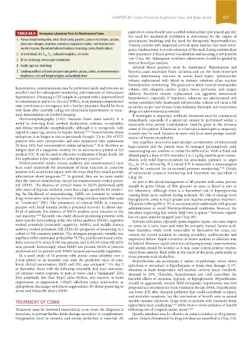
TABLE 88-9 Emergency laboratory Tests for Nonstructural Coma
the need for mechanical ventilation is determined by the degree of
1. Venous blood: hemoglobin, white blood count, platelets, glucose electrolytes, calcium, spontaneous breathing and the need for therapeutic hyperventilation.
blood urea nitrogen, creatinine, osmolality coagulation studies, liver function tests, Trauma patients with suspected cervical spine injuries may need emer-
muscle enzymes, thyroid and adrenal functions, toxicology screen, blood cultures gency tracheostomy to avoid extension of the neck during endotracheal
, carboxyhemoglobin, ammonia tube placement if direct airway visualization and securing is not possible
2. Arterial blood: pH, P CO 2 , P O 2
(see Chap. 46). Subsequent ventilator adjustments should be guided by
3. Urine: toxicology, microscopic examination
arterial blood gas analysis.
4. Gastric aspirate: toxicology Arterial blood pressure must be maintained. Hypotension and
5. Cerebrospinal fluid: cell count and gram stain, protein, glucose, culture, counterimmunoelec- hypoxia cause secondary brain ischemia and are the most important
trophoresis, viral and fungal antigens, and antibody titers factors determining outcome in severe head injury. Intravascular
volume replacement with blood or isotonic solutions often requires
hemodynamic monitoring. The goals are to attain normal intravascular
hypertension, ventriculostomy may be performed safely and provides an volume with adequate cardiac output, tissue perfusion, and oxygen
excellent tool for subsequent monitoring and treatment of intracranial delivery. Excessive volume replacement can aggravate intracranial
hypertension. Obtaining a CSF sample in a patient with a depressed level hypertension, especially if hypotonic solutions are administered and
of consciousness and fever, elevated WBCs, or an immunocompromised serum osmolality falls. Inadequate intravascular volume will cause a fall
state constitutes an emergency and a lumbar puncture should be done in cardiac output and worsen brain ischemia. Inotropic and vasopressor
with haste after carefully ruling out intracranial hypertension or struc- drugs are administered as needed.
tural abnormalities on cerebral imaging. If meningitis is suspected, antibiotic treatment must be commenced
Electroencephalography (EEG) measures brain wave activity. It is immediately, especially if a spinal tap cannot be performed within a
useful in detecting focal cerebral dysfunction, seizures, encephalitis, reasonable time period. Consideration must be given to the immune
and diffuse metabolic encephalopathy, although it is nonspecific with status of the patient. If bacterial or tuberculous meningitis is suspected,
regard to cause (eg, uremia vs hepatic failure). 38,39 Nonconvulsive status steroids may be used. Seizures at onset with fever must prompt consid-
epilepticus is no longer as rare as previously thought. Up to 10% of ICU eration of herpes encephalitis.
patients who were unresponsive and were monitored with continuous Any pupillary anisocoria must prompt consideration of intracranial
24-hour EEG had nonconvulsive status epilepticus. It is therefore an hypertension and the patient must be managed presumptively until
40
integral part of a diagnostic workup for an unconscious patient in the neuroimaging can confirm or refute the differential. With a fixed and
medical ICU. It can be used as objective verification of brain death, but dilated pupil, a dose of 20% mannitol 1 to 1.5 g/kg must be given imme-
this application is less popular in contemporary practice. 41 diately with mild hyperventilation (in atraumatic patients) to target
Evoked-potential studies (visual, auditory, and somatosensory) have to 35 to 40 mm Hg. If a raised ICP is suspected, a neurosurgeon
been used extensively for assessment of brain function in comatose P CO 2 37,44 Details
should be contacted for intracranial pressure monitoring.
patients with acute brain injury, with the hope that they would provide of intracranial pressure monitoring and treatment are described in
information about prognosis. 39,41 In general, they are no more useful Chap. 86.
than the clinical examination, except for somatosensory evoked poten- As part of the initial management of all patients with coma, glucose
tial (SSEP). The absence of cortical waves in SSEPs performed early should be given (50 mL of 50% glucose) as soon as blood is sent to
after onset of hypoxic-ischemic coma has a high specificity for predict- the laboratory. Although there is a theoretical risk of hyperglycemia
ing the likelihood of nonawakening. SSEPs are resistant to sedative causing brain lactic acidosis during ischemia, the risk of damage from
drug intoxication and may be present in drug overdose states that cause hypoglycemic coma is much greater and requires emergency treatment.
an “isoelectric” EEG. The persistence of cortical SSEPs in comatose Thiamine (100 mg IM or IV) is recommended traditionally with glucose
patients with head trauma predicts potential recovery in about one- administration to avoid Wernicke-Korsakoff syndrome; however, the
third of patients; the absence of SSEPs predicts poor outcome in the literature supporting this widely held view is sparse. Seizures, regard-
45
vast majority. 39,42 Recently one study showed promising potential with less of cause, must be stopped (see Chap. 85).
more specific tests looking at the role of late auditory (N100), cognitive Systemic infections, especially gram-negative sepsis, can cause stupor
evoked potentials (mismatch negativity; MMN), and middle latency or coma on a toxic basis and must be promptly treated. Severe acid-
auditory evoked potentials (MLAEPs) for prognosis of awakening in a base disorders, while rarely responsible by themselves for coma, can
cohort of 346 comatose patients. The strongest prognostic variable was worsen the overall situation by causing secondary cardiovascular and
pupillary reflex (estimated probability 79.7%), and the estimated proba- respiratory failure. Rapid correction of severe acidosis or alkalosis may
bility rose to 87% when N100 was present, and to 89.9% when MLAEPs be helpful. However, rapid correction of hyperglycemia, hypernatremia,
were present. Interestingly, when MMN was present, 88.6% of patients and uremia should be avoided as it may cause central pontine myelin-
awakened and no patient with MMN became permanently vegetative. olysis from osmotic fluid shifts in the tracts of the pons, particularly in
In a small study of 34 patients with anoxic coma admitted over a those patients with alcoholism.
2-year period to an intensive care unit, the predictive value of com- Hyperthermia can accompany a variety of pathologic states, either
bined clinical examination, SSEP, and EEG was evaluated. On day 3 infectious or secondary to hypothalamic or brain stem damage. A 1°C
43
or thereafter, those with the following invariably had poor outcomes: elevation in body temperature will increase cerebral tissue metabolic
(a) extensor motor response to pain or worse and a “malignant” EEG demand by 10%. Therefore, hyperthermia can itself exacerbate the
(low amplitude, less than 50 µV, delta rhythm, non-reactive; or burst harmful effects of ischemia, hypoxia, or hypoglycemia. Hyperthermia
suppression; or suppression <20 µV, alfa/theta coma, nonreactive; or should be aggressively treated. Mild therapeutic hypothermia was first
epileptiform discharges with burst-suppression); (b) flexor posturing or proposed as a treatment for brain trauma in the late 1950s. Hypothermia
worse and bilaterally absent SSEPs. can lower ICP, alter chemical pathways that could contribute to injury,
and modulate apoptosis, but the mechanism of benefit seen in animal
TREATMENT OF COMA models remains unknown. Large trials in patients with traumatic brain
injury have been conflicting, 46,47 while there is some evidence of benefit
Treatment must be instituted immediately, even when the diagnosis is following out-of-hospital cardiac arrest. 48,49
uncertain, to prevent further brain damage secondary to complications. Specific antidotes may be effective in coma secondary to drug intoxi-
Oxygenation must be ensured, and airway protection is essential. All cation. Details of treatment for drug overdose are described in Chap. 124.
section06.indd 837 1/23/2015 12:56:24 PM