
Page 1253 - Hall et al (2015) Principles of Critical Care-McGraw-Hill
P. 1253
860 PART 7: Hematologic and Oncologic Disorders
to exhibit severely deficient plasma ADAMTS13 activity. 44,97 These E coli
observations suggest two different mechanisms (autoimmune vs non-
immune) underlying the pathogenesis of TTP caused by ticlopidine Shiga toxin (Stx1 and Stx2)
versus clopidogrel. Patients with ticlopidine-associated TTP often
have severe deficiency of plasma ADAMTS13 activity as a result of Binds to leukocytes
autoantibodies against ADAMTS13 and respond rapidly to plasma
exchange. 44,97 However, patients with clopidogrel-induced TTP rarely
show severe deficiency of plasma ADAMTS13 activity and positive Glomerulus Endothelial cells
anti-ADAMTS13 autoantibodies in their plasma. 43,44
Pregnancy-Associated TTP: Pregnancy has been shown to cause TTP
de novo or act as an inciting factor that triggers an acute episode of B subunit A subunit
TTP in women who have hereditary or acquired deficiency of plasma
ADAMTS13 activity. 23,98,99 In a review of 350 cases by Veyradier et al, UL-VWF Protein synthesis
99
TTP episodes are observed in their first, second, and third trimesters of Tissue factor Apoptosis
pregnancy, as well as during the postpartum period. The time to onset Cytokines Damage of
may suggest the cause: hereditary or acquired. For instance, a woman TM endothelium
with hereditary deficiency of plasma ADAMTS13 activity may develop
TTP in her first pregnancy, primarily in her third trimester, whereas a
woman with acquired deficiency of plasma ADAMTS13 activity due to Platelet adhesion and aggregation
autoantibodies may present her first episode during the first trimester Complement activation
and often after 20 weeks of her gestation. TTP during pregnancy is asso- Fibrin formation
99
ciated with high maternal mortality or long-term morbidity rate. 100-102
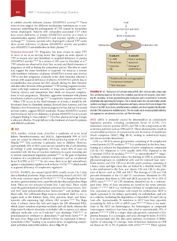
Preterm delivery and intrauterine fetal death are frequent complica- FIGURE 91-2. Mechanism of E coli toxin-induced HUS. After infection with a Shiga toxin
tions of such pregnancies. Therefore, aggressive treatment with plasma (Stx)–producing organism, Stx enters the circulation, possibly via Gb4 receptors. Upon enter-
transfusion or plasma exchange 100-102 is required to improve survival rate. ing the circulation, it binds polymorphonuclear leukocytes and is transferred to vulnerable
When TTP occurs in the third trimester or at term, it should be dif- endothelial cells expressing Gb3 receptors. The A subunit enters the cells and inhibits protein
ferentiated from the Hemolytic anemia, Elevated Liver enzymes, and Low synthesis and triggers endothelial cell apoptosis and injury, whereas the B subunit triggers the
Platelets (HELLP) syndrome. Severe damage to the liver, but not the central release of UL-VWF, tissue factor, proinflammatory cytokines, and downregulates the thrombo-
nervous system, suggests HELLP syndrome. Plasma ADAMTS13 activity modulin (TM) expression in endothelial cells. The end results are to increase platelet adhesion
may be reduced, but severe deficiency of plasma ADAMTS13 activity is not and aggregation, complement activation, and fibrin formation.
a frequent finding in these patients. Therefore, plasma exchange therapy
103
is often not effective. Prompt delivery is the treatment of choice for patients aHUS: aHUS is primarily caused by abnormalities in complement
with HELLP syndrome. regulatory proteins including complement factor H (CFH), CFI,
■ HUS membrane cofactor protein (MCP), and TM or the components in the
activation pathway such as CFB and C3. These abnormalities result in
HUS, another clinical term, describes a syndrome of acute renal uncontrolled activation of complements and formation of membrane
failure, thrombocytopenia, and MAHA. Approximately 90% of HUS attack complexes (MAC) (Fig. 91-3), leading to endothelial injury
cases are caused by infection with a toxin-producing strain of E coli or and microvascular thrombosis.
Shigella. 104,105 This syndrome is primarily seen in children. However, CFH is a 150-kDa serum glycoprotein, consisting of 20 complement
133,134
approximately 10% of HUS cases are not caused by the E coli infection; control protein (CCP) modules. It is synthesized in the liver, func-
its etiology is quite heterogeneous. Of those, nearly 60% of cases are tioning as a cofactor for degradation of active complement component
associated with the loss-of-function mutations in a gene encoding one C3b by CFI. Mutations in CFH, usually (60%-70%) clustered in the
64,137
108,133,135,136
or several complement regulatory proteins 106-112 or the gain-of-function C-terminal CCP19-20 modules, or autoantibodies target-
mutations of a complement activation component such as complement ing these terminal modules reduce the binding of CFH to polyanionic
factor B (CFB) or C3. 113-115 In rare cases, there is an IgG autoantibody glycosaminoglycans on endothelial cells and the exposed base mem-
against a complement regulator such as factor I (CFI), 116-118 which results brane surface, and C3b. CFI is an 88-kDa serine protease, consisting of
in acquired deficiency of complement regulators. a heavy chain and light chain that are linked by a disulfide bond. CFI
is also synthesized in the liver, and cleaves C3b and C4b in the pres-
D+HUS: D+HUS, also named typical HUS, usually occurs 3 to 5 days ence of factors such as CFH and MCP. The cleavage of C3b and C4b
after a diarrheal prodrome. Shiga toxin–producing strain E coli O157 : H7 prevents formation of the C3 and C5 convertases. Mutations in CFI,
is the most common cause of D+HUS. 56,57,119,120 It is thought that bacte- less common than those in CFH and MCP, account for 5% to 12% of
ria infect gastrointestinal tract, cause bloody diarrhea and produce Shiga aHUS cases. 64,112,138,139 All CFI mutations identified are in a heterozy-
toxin. There are two subtypes of toxin (Stx1 1 and Stx2). These toxins gous form. Most of these mutations are found in the serine protease
cross the gastrointestinal epithelium and enter the blood stream. Stx1 domain. 64,112,138,139 MCP is a membrane inhibitor of complement activa-
and Stx2 bind polymorphonuclear leukocytes and perhaps other tion, expressed on most human cells except for erythrocytes. MCP is
121
blood cells. The cell-bound Shiga toxin in circulation finds its way to highly expressed in the kidney, particularly on endothelium, and acts
organs such as the kidneys, brain, liver, pancreas, heart, and hema- as a cofactor, facilitating the degradation of deposited C3b and C4b on
topoietic cells expressing high affinity Gb3 receptor. 122-124 The Shiga host cells. Approximately 20 mutations in MCP have been reported,
toxin A subunit enters the cells and targets the 28S ribosomal RNA, accounting for 10% to 13% of aHUS cases. 64,110,111,140 Most of the muta-
which causes cessation of protein synthesis and cell death, whereas the tions in the MCP are heterozygous, but homozygous or compound
Shiga toxin B subunit stimulates endothelial cells to express and release heterozygous mutations have been described in approximately 25% of
adhesion molecules such as P-selectin and ultralarge VWF 126,127 or patients. 64,110,111,140 CFB is a single-chain glycoprotein composed of five
125
proinflammatory cytokines or chemokines, and tissue factor. 129-131 At protein domains. It is a zymogen, and upon cleavage by factor D (CFD)
128
the same time, Shiga toxin B subunit inhibits the expression of throm- it is incorporated into the alternative pathway convertases (C3bBb),
bomodulin (TM), leading to an acquired defect in regulating comple- capable of catalyzing C3 cleavage. Gain-of-function mutations in CFB
132
ment activation and prothrombotic status (Fig. 91-2). are found in 1% to 3% of patients with aHUS. 64,113,141,142 There appears
section07.indd 860 1/21/2015 7:42:48 AM