
Page 1493 - Hall et al (2015) Principles of Critical Care-McGraw-Hill
P. 1493
1032 PART 9: Gastrointestinal Disorders
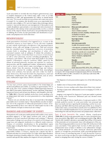
Acute pancreatitis is currently the most frequent gastrointestinal cause TABLE 108-1 Etiology of Acute Pancreatitis
of hospital admissions in the United States with a total of 275,000
admissions in 2009 and approximately $2.2 billion in annual health Toxic Alcohol
1
care costs. The overall mortality among patients with acute pancreatitis Methanol
2
is around 5%, but patients who develop severe acute pancreatitis have Smoking
mortality rates as high as 15% and even higher when multiorgan failure Organophosphates
3
is present. Appropriate intensive care management of these patients and Scorpion bite
a multi-disciplinary approach play a very important role in treatment of Mechanical obstruction/duct Biliary pancreatitis (gallstones)
those patients who develop severe acute pancreatitis. In 2012, a revised damage Biliary sludge
version of the original Atlanta classification was published that focused Parasitic infections (ascariasis)
on defining the severity of acute pancreatitis and classification of pan- Malignancy (pancreatic, ampullary, cholangiocarcinoma)
creatic and peripancreatic fluid collections. 4 Periampullary diverticulum
Abdominal trauma/duct disruption
PATHOPHYSIOLOGY Metabolic Hypertriglyceridemia
Acute pancreatitis is believed to be triggered by an increase in the Hypercalcemia
intraductal pressure or direct injury to acinar cells from metabolic Immune-related Auto-immune pancreatitis
or toxic stimuli which leads to breakdown of the junctional barrier Vasculitis (SLE, polyarteritis nodosa)
between acinar cells and leakage of pancreatic fluid and enzymes Drugs 5-ASA/salicylates, azathioprine/6-MP, didanosine, pent-
into the interstitial space. Intrapancreatic activation of proteolytic amidine, furosemide, tetracyclines, thiazides, estrogen
5
enzymes leads to autophagy and autodigestion of acinar cells.
6
Lysosomal enzymes such as cathepsin B initiate the activation of Infections Viral: mumps, varicella-zoster, coxsackie, HSV, HIV
trypsinogen to trypsin which then leads to activation of more trypsin Bacterial: Mycoplasma, Leptospira, Legionella
as well as other pancreatic enzymes including phospholipase, chy- Parasitic: Toxoplasma, cryptosporidium
motrypsin, and elastase. The acinar tissue death leads to an intense Fungal: Aspergillus
7
systemic inflammatory response syndrome (SIRS) caused by the Miscellaneous Idiopathic
release of activated pancreatic enzymes and mediated by cytokines, Post-ERCP pancreatitis
immunocytes, and the complement system. Inflammatory cytokines Pancreas divisum in some patients
(such as tumor necrosis factor) cause macrophages to migrate into Ischemia
tissues distant from the pancreas, including lungs and kidneys. Genetic mutations in PRSS1, SPINK, CTRC, or CFTR genes
Immunocytes attracted by cytokines released from macrophages Common etiologies highlighted. CFTR, cystic fibrosis transmembrane conductance regulator; CTRC,
release more cytokines, free radicals, and nitric oxide; the result is chymotrypsin C; ERCP, endoscopic retrograde cholangiopancreatography; HIV, human immunodeficiency
tissue destruction, fluid and electrolyte loss, hypotension, renal and virus; HSV, herpes simplex virus; PRSS1, serine protease 1; SLE, systemic lupus erythematosus; SPINK,
pulmonary complications, late septic complications, and, in severe serine protease inhibitor Kazal type 1.
cases, multisystem organ failure (MSOF) and death.
The diagnosis of acute pancreatitis requires two of the following three
ETIOLOGY criteria:
The two most common causes of pancreatitis in the United States are • Sudden onset of characteristic abdominal pain
alcohol and gallstone pancreatitis, accounting for approximately 75% to • Elevation of serum amylase and/or lipase above three times normal
80% of the cases. Other common etiologies include hypertriglyceridemia,
post-ERCP pancreatitis, hypercalcemia, trauma, infections, drug injury, • Findings of pancreatic inflammation noted on imaging (CT, MRI, or
anatomical variants such as pancreas divisum, and idiopathic pancreati- ultrasound)
tis (Table 108-1). Recently, multiple studies have shown smoking to be Imaging is not required for diagnosis of acute pancreatitis in patients
an independent risk factor for acute pancreatitis in a dose-dependent who present with characteristic abdominal pain and elevated serum
manner. 8-10 amylase or lipase.
Patients who are critically ill are also at increased risk of developing Since the original Atlanta classification of acute pancreatitis in 1992,
pancreatitis due to ischemic injury. Hypoperfusion can play an impor- multiple predictive models for acute pancreatitis were proposed and
11
tant role in the progression of mild acute pancreatitis to severe, necro- there was much confusion regarding the terminology used for local com-
tizing pancreatitis in those cases where the initial insult was due to the plications and fluid collections from acute pancreatitis. In an attempt to
more common etiologies in noncritical care setting. 12 address these issues, a revised Atlanta classification was published in 2012
which offers a detailed classification of acute pancreatitis, its severity and
DIAGNOSIS AND ASSESSMENT OF SEVERITY terminology for early and late pancreatic and peripancreatic collections. 4
Most patients with acute pancreatitis present with sudden onset, severe, Types of Acute Pancreatitis: The revised Atlanta classification
persistent epigastric, or right upper quadrant pain, with or without (Table 108-2) divides acute pancreatitis into two types, interstitial edem-
radiation to the back associated with nausea and vomiting. 13,14 Physical atous pancreatitis (Fig. 108-1) and necrotizing pancreatitis (Fig. 108-2).
examination findings vary according to the severity of the disease and Patients with interstitial edematous pancreatitis have diffuse inflammation
range from mild epigastric tenderness to a diffusely tender abdomen. of the pancreatic and peripancreatic tissue with enlargement of the pan-
Presence of ecchymotic discoloration in the periumbilical region creas. Necrotizing pancreatitis is seen in less than 10% of all patients with
(Cullen sign) or along the flanks (Grey Turner sign) suggests retroperi- acute pancreatitis. These patients have necrosis of either pancreatic or
toneal bleed. peripancreatic tissue or both, in addition to the inflammatory changes.
Serum amylase and lipase are both elevated early in the course of acute On contrast-enhanced CT scans, interstitial edematous pancreatitis
pancreatitis (within 4-12 hours). Amylase has a shorter half-life of appears as homogenous enhancement, while pancreatic/peripancreatic
10 hours and returns to normal within 3 to 5 days, while lipase elevations necrosis is seen as nonenhancing areas. Of note, the necrosis of pancre-
last longer, returning to baseline within 8 to 14 days. Serum lipase is more atic tissue can develop over days after onset of abdominal pain and can be
sensitive and specific than amylase for diagnosis of acute pancreatitis. missed on imaging done early during the course of disease. 15,16
section09.indd 1032 1/14/2015 9:27:19 AM