
Page 1496 - Hall et al (2015) Principles of Critical Care-McGraw-Hill
P. 1496
CHAPTER 108: Acute Pancreatitis 1035
■ PANCREATIC AND PERIPANCREATIC COLLECTIONS
The recent Atlanta classification has led to a major change in the clas- Stomach
sification of the pancreatic and peripancreatic fluid collections based
on the presence or absence and duration of solid material in these
collections. The four types of fluid collections as a sequel of acute pan-
4
creatitis include acute fluid collection (AFC), pancreatic pseudocyst (PP)
(Fig. 108-4), acute necrotic collection (ANC), and walled-off necrosis Walled off necrosis
(WON) (Fig. 108-5). All these types of fluid collections have signifi-
cantly different management strategies and hence it is very important to
distinguish one from the others. AFCs develop early in acute interstitial
edematous pancreatitis, are homogenous on contrast-enhanced imaging Pancreas
(no solid debris), do not have well-developed demarcation, and usually Liver
resolve without any intervention. If these persist beyond 4 weeks, they
develop a well-demarcated wall and are known as pseudocysts, which do Spleen
not contain any solid material. Acute necrotic collection (ANC), usually
seen during the first 4 weeks in necrotizing pancreatitis, contains both
fluid and necrotic components and is without a well-demarcated wall.
These can progress to a well-defined encapsulation after 4 weeks, a con-
dition known as walled-off necrosis. Both ANCs and WONs can become
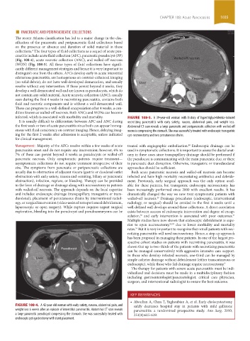
infected, which is associated with morbidity and mortality. FIGURE 108-5. A 39-year-old woman with history of hypertriglyceridemia-induced
It is usually difficult to differentiate between AFC and ANC during necrotizing pancreatitis with early satiety, nausea, abdominal pain, and weight loss.
the first week or two of acute pancreatitis since both can appear homog- Abdominal CT scan reveals a large pancreatic and peripancreatic collection with walled-off
enous with fluid consistency on contrast imaging. Hence, delaying imag- necrosis compressing the stomach. She was successfully treated with endoscopic transgastric
ing for the first 2 weeks after admission is acceptable, unless indicated cyst-necrosectomy and two percutaneous drains.
for clinical management.
Management: Majority of the AFCs resolve within a few weeks of acute treated with angiographic embolization. Endoscopic drainage can be
48
pancreatitis onset and do not require any intervention; however, 6% to used in symptomatic collections. It is important to assess the ductal anat-
7% of these can persist beyond 4 weeks as pseudocysts or walled-off omy in these cases since transpapillary drainage should be performed if
pancreatic necrosis. Only symptomatic patients require treatment— the pseudocyst is communicating with the main pancreatic duct or there
asymptomatic collections do not require treatment irrespective of their is pancreatic duct disruption. Otherwise, transgastric or transduodenal
size. The symptoms from pancreatic or peripancreatic collections are approaches should be sufficient.
usually due to obstruction of adjacent viscera (gastric or duodenal outlet Both acute pancreatic necrosis and walled-off necrosis can become
obstruction with early satiety, nausea and vomiting, biliary or pancreatic infected and have high mortality necessitating antibiotics and debride-
obstruction), infection, rupture, or bleeding. Therapy can be provided ment. Previously, early surgical approach was the only option avail-
in the form of drainage or drainage along with necrosectomy in patients able for these patients, but transgastric endoscopic necrosectomy has
with walled-off necrosis. The approach depends on the local expertise been increasingly performed since 2000 with excellent results. It has
and includes: endoscopic drainage (transpapillary, transgastric or trans- dramatically changed the way we now treat symptomatic patients with
duodenal), placement of percutaneous drains by interventional radiol- walled-off necrosis. Drainage procedures (endoscopic, interventional
49
ogy, or surgical intervention (video-assisted retroperitoneal debridement, radiology, or surgical) should be avoided in the first 4 weeks until a
laparoscopic or open surgery). While rupture requires urgent surgical well-defined wall develops around these collections. A direct correlation
exploration, bleeding into the pseudocyst and pseudoaneurysms can be exists between success of endoscopic intervention and degree of encap-
sulation, and early intervention is associated with poor outcomes.
51
50
Multiple studies have now shown that endoscopic debridement is supe-
rior to open necrosectomy, 52,53 due to lower morbidity and mortality
rates. But it is very important to recognize that not all patients with nec-
54
rotizing pancreatitis will need necrosectomy. Hence, a step-up approach
has been proposed in managing these patients. In one of the largest pro-
spective cohort studies on patients with necrotizing pancreatitis, it was
shown that up to two-thirds of the patients with necrotizing pancreatitis
Pseudocyst
can be managed conservatively with aggressive intensive care support.
In those who develop infected necrosis, one-third can be managed by
Liver
simple catheter drainage without debridement (either transcutaneous or
endoscopic), while those who fail drainage require necrosectomy. 3
The therapy for patients with severe acute pancreatitis must be indi-
vidualized and decisions must be made in a multidisciplinary fashion
Spleen
including gastroenterologist/pancreatologist, critical care physician,
surgeon, and interventional radiologist to ensure the best outcome.
KEY REFERENCES
• Aboulian A, Chan T, Yaghoubian A, et al. Early cholecystectomy
FIGURE 108-4. A 42-year-old woman with early satiety, nausea, abdominal pain, and safely decreases hospital stay in patients with mild gallstone
weight loss 6 weeks after an episode of interstitial pancreatitis. Abdominal CT scan reveals pancreatitis: a randomized prospective study. Ann Surg. 2010;
a large pancreatic pseudocyst compressing the stomach. She was successfully treated with 251(4):615-619.
endoscopic cyst-gastrostomy with stent placement.
section09.indd 1035 1/14/2015 9:27:24 AM