
Page 1498 - Hall et al (2015) Principles of Critical Care-McGraw-Hill
P. 1498
CHAPTER 109: Mesenteric Ischemia 1037
TABLE 109-1 Potential Causes of Acute Mesenteric Ischemia
Occlusive Disease
Embolism Cardiac diseases (atrial fibrillation, post-MI, valvular disease,
SBE, dilated left ventricle, myxoma)
Extracardiac arterial diseases Celiac branches
Arterial thrombosis Acute on chronic atherosclerosis
Low cardiac output states
Intrinsic arterial dis- Occlusive atherosclerosis Arch of riolan
eases Aortic dissection (type A or B)
Atherosclerotic aneurysm
Arteritis and autoimmune diseases
Fibromuscular dysplasia
Venous thrombosis Thrombophilia
Extrinsic compression
Iatrogenic After aortoiliac surgery
Catheter related (dissection, embolism)
Irradiation arteritis
Vasoconstrictive agents (epinephrine, norepinephrine,
dopamine)
Distal SMA branches
Trauma Penetrating
Blunt (including deceleration injuries)
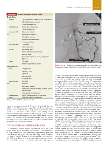
FIGURE 109-1. Late IMA injection demonstrating the IMA to the arch of Riolan, filling
Nonocclusive Disease
the stump of the SMA with its distal branches. Also note filling of hepatic and splenic branches.
Shock Cardiogenic shock
Hypovolemic shock
Septic shock
times of stress or increased demand. This preferential shunting of blood
Neurogenic shock
to vital organs, if acute or severe, can “sacrifice” the mesenteric circula-
Anaphylactic shock tion, leading to low-flow and ischemic injury. The more metabolically
Low cardiac output Heart failure active mucosal layer receives 70% of blood flow, only 30% supplying the
states Arrhythmia muscularis and serosal layers, placing the intestinal mucosa at greatest
risk from ischemic injury. Within the intestinal villus, passive exchange
6
Acute coronary syndromes of oxygen typically occurs between the afferent arteriole and efferent
Miscellaneous : peritonitis ,pancreatitis, post CABG, ESRD on venule, effectively bypassing the capillary network at the villus tip, a phe-
peritoneal dialysis nomenon called oxygen countercurrent exchange. In health, high partial
Pseudocoarctation (aortic dissection) pressures of oxygen ensure that the metabolic needs of the villus mucosa
are met despite this shunting, but in deoxygenated states this shunting
Pharmacologically Digitalis, vasoactive substances (catecholamines, adversely affects oxygen delivery to the mucosal tip, making it most vul-
induced somatostatin analogues, etc.), ergotism nerable to ischemic injury. A number of extrinsic and intrinsic factors
MI, myocardial infarction; SBE, subacute bacterial endocarditis. regulate the mesenteric blood flow, leading to a complex interaction
between neural, hormonal, and paracrine effectors that regulate the vas-
cular smooth muscle tone in the mesenteric bed and control local blood
vessels to the marginal artery of Drummond (a collateral that runs flow. Vasoactive mediators alter the vascular smooth muscle (VSM) tone
within 1-2 cm of the mesenteric edge of the bowel) and via the arch of multiple small afferent arterioles, collectively known as resistance
of Riolan. Vessel caliber decreases progressively from the main aortic vessels, changing their cross-sectional area and blood flow (Table 109-2).
branch to the mesenteric vascular arcades distally, ending with an The interaction between extracellular agonist (first messenger) and
extensive communicating submucosal vascular plexus. This collateral VSM receptor leads to accumulation of intracellular second messengers,
network explains why many individuals may tolerate chronic occlusion such as cyclic adenosine monophosphate (cAMP), Ca , and cyclic
2+
of one or two mesenteric vessels without symptoms, but it also explains guanosine monophosphate (cGMP). These second messengers directly
2+
why acute-on-chronic occlusion of one additional branch may lead to a or indirectly alter the cytosolic concentration of Ca and dictate
7
catastrophic loss of intestinal perfusion. whether VSM contracts. A functioning cardiovascular system is essen-
tial because the mesenteric circulation is often sacrificed to maintain
CONTROL OF THE MESENTERIC CIRCULATION blood flow to vital organs at times of detrimental alterations in cardiac
output, blood volume, or arterial blood pressure. Increased sympathetic
The mesenteric circulation receives approximately 20% to 30% of the nervous activity associated with cardiogenic, septic, or hypovolemic
6
cardiac output at rest, which may increase by up to 50% after meals. shock can further compound flow-related ischemia by inducing intense
As such, the mesenteric circulation receives approximately three times vasoconstriction within the mesenteric bed. High-volume hemorrhage
more blood per unit weight than most other body tissues. This blood (>35% of blood volume) leads to disproportionate visceral vaso-
flow is partly to satisfy the absorptive function of the intestine and per- constriction compared with the reduction in cardiac output. When
8
fuse the liver via the portal vein, but it also represents a reservoir func- activated postganglionic sympathetic nerves fibers release norepineph-
tion from which blood can be mobilized to other sites (vital organs) at rine, stimulating α -adrenergic receptors on VSM of precapillary arterioles
2
section09.indd 1037 1/14/2015 9:27:26 AM