
Page 1502 - Hall et al (2015) Principles of Critical Care-McGraw-Hill
P. 1502
CHAPTER 109: Mesenteric Ischemia 1041
(>5 mm) bowel wall and signs of ileus (distended bowel loops and methods. Vascular findings on CT that contribute to the diagnosis of
hypoperistalsis). In advanced cases (mesenteric infarction), ultrasound mesenteric ischemia include arterial stenosis, embolus visualization,
may show intraperitoneal fluid, pneumatosis, or intrahepatic portal arterial aneurysm with thrombus, thrombosis of mesenteric vessels,
venous gas. However, ultrasonography is operator-dependent and may arterial dissection, and mesenteric vein thrombosis. Nonvascular CT
be confounded by the degree of intraluminal intestinal gas or by obe- findings include bowel wall thickening, hypoperfusion and hypoattenu-
sity. For patients with suspected chronic mesenteric ischemia, duplex ation, bowel dilation, bowel wall hemorrhage, mesenteric fat stranding,
ultrasonography has become the investigation of choice by many. A pneumatosis intestinalis, and portal venous gas. Alternatives to iodinated
67
31
peak systolic velocity of greater than 275 cm/s in the SMA or greater contrast agents, such as gadolinium, may be used for patients with a
than 200 cm/s in the celiac artery or no flow signal (in the SMA or celiac contraindication to standard contrast material. Ischemic bowel wall
artery) predicts a stenosis of greater than 70% with a sensitivity of 89% typically is seen as thin and poorly enhanced, more prominent on the
and a specificity of 92%. antimesenteric border in NOMI. Air may be seen in the bowel wall or
Following successful bypass or revascularization, postoperative the portal vein. Reperfused bowel segments appear enlarged and edema-
duplex surveillance can be used to assess the patency of bypass grafts to tous, often with increased mucosal and submucosal enhancement due
mesenteric arteries. Although the inflow arterial peak systolic velocity to interstitial extravasation of contrast material. With the increased
32
may be higher for retrograde bypasses, the anastomotic and midgraft availability of modern multislice helical CT scanners, rapidly acquired
velocities are not significantly affected by the orientation of the graft. 68 high-quality CTA images of the main mesenteric trunks and small col-
■ CT SCAN AND CT-ANGIOGRAM laterals are possible. Compared with mesenteric angiography, CTA
57
may prove safer, cheaper, and better tolerated and result in decreased
CT scanning has become the radiologic investigation of choice for acute radiation exposure for both patient and staff. When combining vascular
abdominal pain. Oral contrast enhancement may give better luminal imaging and bowel wall appearance, the specificity is reported at 94%
definition but is not used by many centers, especially in the emergency and sensitivity of 96%. 67
further elaborates the major vessel flow and tissue perfusion (Fig. 109-3). ■ MRI AND MR-ANGIOGRAPHY (MRA)
situation. Intravenous contrast enhancement (CT-angiography, CTA)
Both arterial and venous phases can be performed for additional The superior soft tissue definition and non-contrast-enhanced angio-
information. All three mesenteric vessels can be identified and patency graphic ability of MRI give it potential advantages in patients with an
66
assessed by their contrast content. Degree of stenosis and calcification acute abdomen. Recent advances in MRA technology and the use of
33
are useful in planning arterial interventions either by catheter or surgical contrast-enhanced (CE) techniques have shortened acquisition times and
reduced the impact of motion artifacts, which previously had restricted
its use. MRA may prove to be the investigation of choice in chronic
34
A mesenteric ischemia, where it has been suggested that CE-MRA is supe-
rior to digital subtraction angiography for simultaneous exploration of
the abdominal aorta and its major branches, and it can be coupled with
measurements of flow and assessment of surrounding soft tissues.
35
However, at present, its use in emergency situations is reduced due to
longer examination times and motion artifacts due to patient movement. 67
■ ANGIOGRAPHY
Mesenteric angiography remains the most specific test for the diagnosis
of mesenteric ischemia, giving objective, reproducible evidence and in
some instances providing therapeutic catheter-based options. Not only
36
does it identify the location of the flow-limiting lesion, but it also may
give information about distal runoff and the extent of collateralization,
allowing the most appropriate therapy to be planned. However, it can-
not discern whether or not intestinal infarction has occurred, does not
provide details of the bowel wall changes and therefore, treatment always
must be planned with full assessment of clinical and laboratory param-
B eters, especially if a catheter-based therapy is planned. 37
Thrombotic occlusion usually occurs on a background of chronic
atherosclerotic disease, where lesions tend to be at or near the ostium,
producing an abrupt cutoff of contrast (Fig. 109-4). The chronicity of the
atherosclerotic process may be indicated by the presence of multivessel
disease or the presence of large collaterals refilling the branch territories
distally. One must consider the clinical findings carefully because
chronic mesenteric occlusions are seen in up to 60% of the octogenarian
population but cause symptoms in only 5%.
Mesenteric artery emboli present as sharp, rounded filling defects
with a typical meniscus sign. Distal vessels may refill through collateral
vessels. Emboli typically lodge at the sites of vessel narrowing, such as
the origin or a bifurcation. The SMA is affected most often because it
is a relatively large vessel with high flow, and its orientation encourages
antegrade entry of the embolus. SMA emboli typically occlude the vessel
just distal to the middle colic artery origin (Fig. 109-5).
Nonocclusive mesenteric ischemia is typified by diffuse narrowing of
the mesenteric artery and its branches, alternating areas of narrowing
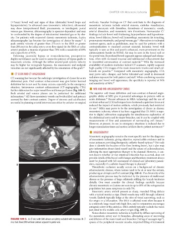
FIGURE 109-3. A. CT slice with SMA almost completely occluded with thrombus. B. CT and dilation of the main trunk and branches (“string of sausages sign”),
slice that shows SMA without contrast adjacent to portal vein. spasm of the peripheral vascular arcades, impaired filling of intramural
section09.indd 1041 1/14/2015 9:27:28 AM