
Page 1499 - Hall et al (2015) Principles of Critical Care-McGraw-Hill
P. 1499
1038 PART 9: Gastrointestinal Disorders
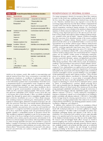
TABLE 109-2 Potential Vasoactive Mediators of the Enteric Circulation PATHOPHYSIOLOGY OF INTESTINAL ISCHEMIA
Mediator Vasoconstrictors Vasodilators The unique arrangement whereby the mesenteric blood flow continues
Neural ↑ Sympathetic tone (adrenergic) ↓ Sympathetic tone (cholinergic) in series via the portal vein, supplying much of the metabolic need of
the liver, means that oxygen extraction by intestinal tissue remains low
↓ Parasympathetic tone ↑ Parasympathetic tone at rest. Therefore, intestinal tissues may respond to reduced flow by
Neuropeptide Y Substance P increasing oxygen extraction from the blood without a requirement for
Vasoactive intestinal peptide (VIP) increased blood flow, but this deprives the liver of some of its portal-
derived oxygenation, resulting in hepatic ischemia. On a biochemical
Calcitonin gene-related peptide (CGRP-α)
and microscopic level, acute ischemia of the intestine is characterized by
Humoral Catecholamines (except liver Catecholamines (only liver and muscle) depletion of cellular adenosine triphosphate (ATP). This leads to failure
and muscle) of the key energy-dependent processes in the cells (in particular mem-
Angiotensin II Histamine brane ATPase pumps) and results in cellular swelling, membrane disrup-
tion, metabolic arrest, and cell death. Hypoxic conditions themselves
11
Vasopressin Bradykinin
favor the conversion of the abundant amounts of the enzyme xanthine
Serotonin Activated complement (C3a, C5a) dehydrogenase to xanthine oxidase. Hypoxia results in accumulating
Activated complement (C5a) Adrenomedullin adenosine levels which can be metabolized further to inosine and then
Paracrine/ Endothelin-1 (VSM cells) Endothelium-derived relaxing factor (EDRF) hypoxanthine but no further without oxygen. With the reintroduction
of oxygen on reperfusion, xanthine oxidase converts hypoxanthine into
autocrine Platelet-activating factor xanthine, producing superoxide (reperfusion tissue injury ). Oxygen
12
Constrictor prostaglandins (F α) Endothelium-derived hyperpolarizing free-radicals (OFRs), including superoxide anions (O ), hydrogen per-
−
2 2
−
factor oxide (H O ), and hydroxyl radicals (OH ), arise as by-products of the
2
2
xanthine-xanthine oxidase system, the mitochondrial electron transport
Dilator prostaglandins (I or prostacyclin) system, and the NADPH oxidase system of infiltrating neutrophils.
2
Endothelin-1 (endothelial cells) Endogenous defenses against oxidative injury, including intracellular
Metabolic ↑ P O 2 ↓ P O 2 enzymes (superoxide dismutase, catalase, and glutathione peroxidase)
and vitamins (C and E), can be overwhelmed by the production of
↓ P CO 2 ↑ P CO 2 OFRs during reperfusion. These OFRs directly attack cell membranes,
↑ pH ↓ pH resulting in lipid peroxidation and synthesis of lipid mediators (throm-
↓ Metabolites (K , lactate, ↑ Metabolites boxane A , leukotriene B ) and chemotactic peptides (complement
+
4
2
adenosine, etc) component C5a), which promote neutrophil activation and chemotaxis,
augmenting the inflammatory component. Sequestration of circulating
neutrophils in the microvessels of the intestine begins with constitutively
expressed neutrophil L-selectin receptors binding to the postcapillary
(which are the resistance vessels), this results in vasoconstriction and venule endothelial P-selectin and E-selectin receptors. This is the first
13
reduced intestinal blood flow. Some compensation is provided by the phase of neutrophil adhesion, manifested by neutrophil rolling along
simultaneous stimulation of vasodilatory β -adrenergic receptors on the venular luminal surface. A second phase of neutrophil adhesion
2
VSM by norepinephrine. However, the net effect is a rapid reduction and influx into the tissue occurs after upregulation of the CD11/CD18
9
in flow followed by a gradual return to prestimulation blood flow levels, integrins and shedding of L-selectin from the neutrophil surface, caused
a phenomenon known as autoregulatory escape. Exogenously admin- by release of proinflammatory cytokines, platelet-activating factor
istered norepinephrine similarly can cause nonocclusive mesenteric (PAF), and eicosanoids from the damaged endothelial cell. This halts
ischemia (NOMI). Parasympathetic nerves induce vasodilation due to neutrophil rolling in the blood vessels of the intestine and by providing
the effect of specific neurotransmitters acetylcholine (ACh, increases strong neutrophil-endothelial cell adhesion through the interaction of
cGMP and NO), vasoactive intestinal peptide (VIP, increases cAMP), the CD11b/CD18 integrin with the endothelial receptor, intercellular
and adenosine triphosphate (ATP). Primary sensory nerves also play adhesion molecule 1 (ICAM-1). Finally, neutrophils transmigrate in
14
a role because their activity through C fibers can inhibit sympathetic response to chemotactic stimuli (such as interleukin-8) and binding by
impulse flow through the spinal cord or sympathetic ganglia. Indeed, platelet-endothelial cell adhesion molecule 1 (PECAM-1). Neutrophils
direct antidromic vasodilation can occur as C fibers release neuropep- further damage tissues by releasing superoxide anions via the NADPH
tides, including substance P, calcitonin gene–related peptide, and VIP, in oxidase system, secreting myeloperoxidase that catalyzes the production
response to luminal signals. Humoral (endocrine) messengers also can of hypochlorous acid (HOCL), and releasing granular enzymes, includ-
affect the mesenteric blood flow, most notably adrenal catecholamines ing elastase, collagenase, and cathepsin G.
in states of systemic stress, shock, or secreting tumor (pheochromo- In addition, many capillaries fail to perfuse on reinstitution of blood
cytoma). Similarly vasoconstrictive effects are seen with renal-derived flow, the no-reflow phenomenon, due to loss of local autoregulation com-
angiotensin II in congestive heart failure and pituitary-derived vasopres- bined with mechanical obstruction of narrowed capillaries by enlarged
sin in shock, respectively. Ingested meals and gastrointestinal luminal adherent neutrophils. No-reflow results in incomplete and patchy tissue
15
peptides stimulate a postprandial increase in mesenteric blood flow. reperfusion, prolonging hypoxia and exposure of the tissues to toxic
This vasodilatory response is regulated, in part, by the increased meta- metabolites. On a microscopic level, complete ischemia causes detectable
bolic activity in the tissues, leading to local accumulation of vasoactive injury to the superficial part of the mucosa (the villi) within 20 minutes.
metabolites such as amines, peptides, prostanoids, and adenosine in the As the duration of ischemia increases, the villus loss can be complete before
presence of a reduced oxygen concentration. Adenosine itself, increasing the muscular layers are damaged. Patchy cellular necrosis can extend to
as a result of energy-dependent processes, may vasodilate in a paracrine mucosal sloughing seen clinically as bloody diarrhea. Ultimately, there is
fashion. A number of hormones are also released in response to luminal transmural infarction, perforation, and septic peritonitis. Prior to reperfu-
contents, including gastrin, cholecystokinin, and secretin, with net vaso- sion, metabolic products such as lactate and other acids are not absorbed
dilatory effects. Somatostatin, released by the mucosal D cells, induces via the negligible portal circulation but only through the peritoneal
10
mucosal vasoconstriction, and synthetic analogs (eg, octreotide) can surfaces, accounting for the lack of acidosis or elevated serum lactate
induce ischemia. commonly observed in patients presenting with acute mesenteric ischemia.
section09.indd 1038 1/14/2015 9:27:26 AM