
Page 1530 - Hall et al (2015) Principles of Critical Care-McGraw-Hill
P. 1530
CHAPTER 110: Special Considerations in the Surgical Patient 1049
in the surgical patient is the early identification of occult sources of lung is thought to occur within 5 minutes after induction of general
sepsis, aggressive investigation for abdominal causes of sepsis, and the anesthesia.
provision of adequate drainage and treatment of septic foci, particularly Shunting results from continued perfusion of nonventilated lung
within the abdomen. units, and the major cause of this imbalance in the surgical patient is
Although both increased microvascular hydrostatic pressure and perioperative atelectasis, although alveolar edema from fluid overload or
pulmonary capillary permeability are important factors in the elabora- capillary leakage could also result in an increase in shunting.
tion of extravascular lung water, manipulation of the microvascular
pressure (by the use of vasoactive agents and regulation of the state of Age, Position, and Airway Closure: Most surgical patients undergo proce-
hydration) is the most direct means of altering pulmonary edema in dures in the supine position, and we are operating increasingly on elderly
the surgical patient. A search for a septic focus in the surgical patient is patients. Also, one of the major effects of surgery is the pain resulting
crucial whenever there is evidence of increased capillary permeability. from surgical incisions. Body position, incisional pain, and age all affect
Control of capillary permeability can then be achieved, although only the relationship between the functional residual capacity (FRC) and the
indirectly, by treating the source of sepsis, which may be surgically closing volume. The FRC has been considered the most important index
approachable. The link between sepsis and capillary permeability is thus of mechanical abnormality in the lung because it represents the balance
broken, and the capillary permeability lesion is allowed to resolve with of opposing forces on the rib cage at resting lung volume. The closing
time; its resolution is accompanied by improvement in perioperative volume is the volume of the lungs at which airway closure begins. When
respiratory failure. Until the permeability corrects itself, reduction of FRC exceeds closing volume, lower airway patency is maintained, while
39
PAWP to the lowest level associated with adequate peripheral perfusion airway closure begins when the FRC falls below the closing volume.
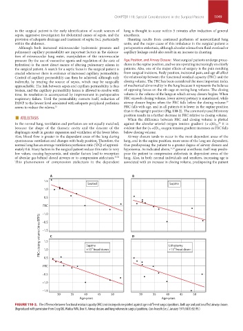
seems to reduce the edema. 32 FRC falls with age, and in all patients it is lower in the supine position
than in the upright position (Fig. 110-2). The commonly used lithotomy
■ ATELECTASIS position results in a further decrease in FRC relative to closing volume.
When the difference between FRC and closing volume is plotted
In the normal lung, ventilation and perfusion are not equally matched, against the alveolar-arterial oxygen tension gradient (a-a)D O 2 , it is
40
because the shape of the thoracic cavity and the descent of the evident that the (a-a)D O 2 oxygen tension gradient increases as FRC falls
diaphragm result in greater expansion and ventilation of the lower lobes. below closing volume.
Also, blood flow is greater in the dependent areas of the lung during Airway closure tends to occur in the most dependent areas of the
spontaneous ventilation and changes with body position. Therefore, the lung, and in the supine position, more areas of the lung are dependent,
x x
normal lung has an average ventilation:perfusion ratio (V /Q) of approxi- thus predisposing the patient to a greater degree of airway closure and
mately 0.8. Many factors in the surgical patient reduce this ratio to very hypoxemia. As indicated above, 37,38 general anesthesia itself may predis-
low values, causing hypoxemia, and similar factors lead to resorption pose the patient to compression atelectasis in dependent areas of the
of alveolar gas behind closed airways or to compression atelectasis. 37,38 lung. Also, in both normal individuals and smokers, increasing age is
This phenomenon of compression atelectasis in the dependent associated with an increase in closing volume, predisposing the patient
A B
1.5
Seated Supine
1.0
0.5
FRC
–1.0
FRC-closing volume (litres BTPS) –1.5 C Supine D Lithotomy
–0.5
1.5
º
º
1.0
0.5 +15 head down +15 head down
FRC
–0.5
–1.0
–1.5
30 35 40 45 50 30 35 40 45 50
Age-years Age-years
FIGURE 110-2. The difference between functional residual capacity (FRC) and closing volume plotted against age in different surgical positions. Both age and position affect airway closure.
(Reproduced with permission from Craig DB, Wahba WM, Don H. Airway closure and lung volumes in surgical positions. Can Anaesth Soc J. January 1971;18(1):92-99.)
section10.indd 1049 1/20/2015 9:19:30 AM