
Page 1554 - Hall et al (2015) Principles of Critical Care-McGraw-Hill
P. 1554
CHAPTER 112: Principles of Postoperative Critical Care 1073
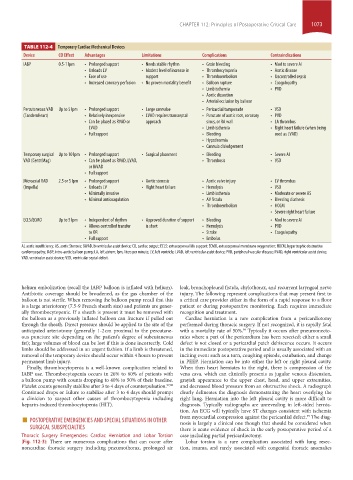
TABLE 112-4 Temporary Cardiac Mechanical Devices
Device CO Effect Advantages Limitations Complications Contraindications
IABP 0.5-1 lpm • Prolonged support • Needs stable rhythm • Groin bleeding • Mod to severe AI
• Unloads LV • Modest level of increase in • Thrombocytopenia • Aortic disease
• Ease of use support • Thromboembolism • Uncontrolled sepsis
• Increased coronary perfusion • No proven mortality benefit • Balloon rupture • Coagulopathy
• Limb ischemia • PVD
• Aortic dissection
• Arterial occlusion by balloon
Percutaneous VAD Up to 5 lpm • Prolonged support • Large cannulae • Pericardial tamponade • VSD
(TandemHeart) • Relatively inexpensive • LVAD requires transseptal • Puncture of aortic root, coronary • PVD
• Can be placed as RVAD or approach sinus, or RA wall • LA thrombus
LVAD • Limb ischemia • Right heart failure (when being
• Full support • Bleeding used as LVAD)
• Hypothermia
• Cannula dislodgement
Temporary surgical Up to 10 lpm • Prolonged support • Surgical placement • Bleeding • Severe AI
VAD (CentriMag) • Can be placed as RVAD, LVAD, • Thrombosis • VSD
or BiVAD
• Full support
Microaxial VAD 2.5 or 5 lpm • Prolonged support • Aortic stenosis • Aortic valve injury • LV thrombus
(Impella) • Unloads LV • Right heart failure • Hemolysis • VSD
• Minimally invasive • Limb ischemia • Moderate or severe AS
• Minimal anticoagulation • AV fistula • Bleeding diathesis
• Thromboembolism • HOCM
• Severe right heart failure
ECLS/ECMO Up to 5 lpm • Independent of rhythm • Approved duration of support • Bleeding • Mod to severe AI
• Allows controlled transfer is short • Hemolysis • PVD
to OR • Stroke • Coagulopathy
• Full support • Embolus
AI, aortic insufficiency; AS, aortic Stenosis; BiVAD, biventricular assist device; CO, cardiac output; ECLS: extracorporeal life support; ECMO, extracorporeal membrane oxygenation; HOCM, hypertrophic obstructive
cardiomyopathy; IABP, intra-aortic balloon pump; LA, left atrium; lpm, liters per minute; LV, left ventricle; LVAD, left ventricular assist device; PVD, peripheral vascular disease; RVAD, right ventricular assist device;
VAD, ventricular assist device; VSD, ventricular septal defect.
helium embolization (recall the IABP balloon is inflated with helium). leak, bronchopleural fistula, chylothorax, and recurrent laryngeal nerve
Antibiotic coverage should be broadened, as the gas chamber of the injury. The following represent complications that may present first to
balloon is not sterile. When removing the balloon pump recall that this a critical care provider either in the form of a rapid response to a floor
is a large arteriotomy (7.5-9 French sheath size) and patients are gener- patient or during postoperative monitoring. Each requires immediate
ally thrombocytopenic. If a sheath is present it must be removed with recognition and treatment.
the balloon as a previously inflated balloon can fracture if pulled out Cardiac herniation is a rare complication from a pericardiotomy
through the sheath. Direct pressure should be applied to the site of the performed during thoracic surgery. If not recognized, it is rapidly fatal
anticipated arteriotomy (generally 1-2 cm proximal to the percutane- with a mortality rate of 50%. Typically it occurs after pneumonecto-
99
ous puncture site depending on the patient’s degree of subcutaneous mies where a part of the pericardium has been resected; either a small
fat); large volumes of blood can be lost if this is done incorrectly. Cold defect is not closed or a pericardial patch dehiscence occurs. It occurs
limbs should be addressed in an urgent fashion. If a limb is threatened, in the immediate postoperative period and is usually associated with an
removal of the temporary device should occur within 4 hours to prevent inciting event such as a turn, coughing episode, extubation, and change
permanent limb injury. in PEEP. Herniation can be into either the left or right pleural cavity.
Finally, thrombocytopenia is a well-known complication related to When then heart herniates to the right, there is compression of the
IABP use. Thrombocytopenia occurs in 26% to 60% of patients with vena cava, which can clinically presents as jugular venous distension,
a balloon pump with counts dropping to 40% to 50% of their baseline. grayish appearance to the upper chest, head, and upper extremities,
Platelet counts generally stabilize after 3 to 4 days of counterpulsation. 97,98 and decreased blood pressure from an obstructive shock. A radiograph
Continued drops or failure to stabilize after 3 to 4 days should prompt clearly delineates the diagnosis demonstrating the heart overlying the
a clinician to suspect other causes of thrombocytopenia including right lung. Herniation into the left pleural cavity is more difficult to
heparin-induced thrombocytopenia (HIT). diagnosis. Typically radiographs are unrevealing in left-sided hernia-
tion. An ECG will typically have ST changes consistent with ischemia
■ POSTOPERATIVE EMERGENCIES AND SPECIAL SITUATIONS IN OTHER from myocardial compression against the pericardial defect. The diag-
99
SURGICAL SUBSPECIALTIES nosis is largely a clinical one though that should be considered when
there is acute evidence of shock in the early postoperative period of a
Thoracic Surgery Emergencies: Cardiac Herniation and Lobar Torsion case including partial pericardiectomy.
(Fig. 112-3): There are numerous complications that can occur after Lobar torsion is a rare complication associated with lung resec-
noncardiac thoracic surgery including pneumothorax, prolonged air tion, trauma, and rarely associated with congenital thoracic anomalies
section10.indd 1073 1/20/2015 9:19:45 AM