
Page 1556 - Hall et al (2015) Principles of Critical Care-McGraw-Hill
P. 1556
CHAPTER 112: Principles of Postoperative Critical Care 1075
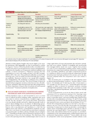
TABLE 112-5 Neurological injury after Carotid Revascularization
Atheroembolic Thrombotic Hypoperfusion Cerebral Hyperperfusion
Mechanism Atherosclerotic plaques become Disruption of intima creates a Drop in CPP usually related to Unopposed flow to areas of prior
dislodged during the procedure by prothrombotic state that leads to decreased MAP loss of cerebral vascular bed
either spontaneous rupture or iatrogenic thrombus formation at the site of autoregulation
manipulation and distally embolize revascularization
Frequency of 54% (Along with thrombotic) 54% (Along with atheroembolic) 19% 15%
occurrence
CEA vs CAS Theoretically less common in CAS Little research done, some suggestion for Theoretically lower risk in CAS for No theoretical variation; known to
due to filter protective devices and risk of late (<3 months) in-stent stenosis patients with contralateral occlusion occur with both
trapping of plaques under stent with stopping of antiplatelet therapy due to decreased ipsilateral
occlusion/clamp time
Diagnostic testing TCD TCD TCD, cerebral oximetry, EEG TCD, dynamic susceptibility MRI,
SPECT, cerebral angiography
Symptoms Acute neurological change Acute neurological change Reduction in flow readings of Spectrum from unilateral headache
intraoperative monitors, to seizures to transient neurological
intraoperative neurological changes symptoms to cerebral hemorrhage
with clamping on awake patient
Timing of presentation Intraoperative or first several hours Immediately postoperatively to first Intraoperatively Within 36 hours postoperatively
postoperatively several hours postoperatively
Prevention/treatment Precise surgical techniques; embolic Possibly antiplatelet therapy (ASA), use Maintenance of intraoperative Maintain SBP <120-140 with
protection devices; possibly closed- of dextran perfusion: shunting, permissive clonidine or ß-blockers, avoid CCB
cell stents hypertension; avoidance of and nitrates
general anesthesia
Immediate reexploration and
thrombectomy
CAS, carotid artery stenting; CCB, calcium channel blockers; CEA, carotid endarterectomy; CPP, cerebral perfusion pressure; MAP, mean arterial pressure; MRI, magnetic resonance imaging; SPECT, single-photon
emission computed tomography; SBP, systolic blood pressure; TCD, transcranial Doppler.
clinician to obtain a hepatic Doppler with vascular duplex of the vascu- a large, rapid release of lactate and potassium that can lead to arrhyth-
lar anastomoses. MRI angiography can then be performed to confirm mias and severe acidosis. Washout of myoglobin and microthrombi
the diagnosis. Presence of thrombus should result in immediate throm- from damaged skeletal muscle also occurs. Together these products
bectomy in an effort to prevent graft loss and need for retransplantation. can result in local complications including massive edema requiring
Rejection after transplant can be hyperacute (antibody mediated), acute fasciotomies for compartment syndrome and systemic complications of
(cellular), or chronic (ductopenic). Hyperacute rejection after liver shock, renal failure, arrhythmias, and death. Compartment syndrome
103
transplantation is a rare event usually associated with ABO incompat- is a condition in which an enclosed myofascial compartment develops
ibility in 60% of the cases. Other preformed antibodies account for the elevated pressure that limits perfusion to the muscles, nerves, fat, skin,
remaining cases. Cases are usually associated with high titers of antido- and bone contained within the compartment and is also covered in
nor antibodies. Hyperacute rejection is rare among ABO-compatible chapter 122.
liver transplantations. The mechanism is preformed donor antibodies Time of ischemia prior to reperfusion is the most important factor
causing graft loss within a few days. The clinical findings of hyperacute to determining postreperfusion syndrome. Treatment before 12 hours
rejection include progressive elevation of liver function tests, thrombo- leads to 19% mortality and 7% limb loss, but after 12 hours leads to a
cytopenia, and hepatic failure during the first days after transplantation, 31% mortality and 22% limb loss risk. Several alternatives have been
104
but biliary obstruction or hepatic artery thrombosis will not be present suggested to reduce or prevent the reperfusion syndrome, including
an ultrasound. Treatment is primarily retransplantation. controlled limb reperfusion (rather than acute restoration to normal
103
blood flow), administered hypothermia, and femoral venous drainage
■ VASCULAR SURGERY EMERGENCIES: POSTREPERFUSION SYNDROME of affected limb. 104
Carotid revascularization occurs in the form of either carotid end-
AND NEUROLOGIC INJURY AFTER CAROTID REVASCULARIZATION arterectomy (CEA) or carotid artery stenting (CAS). Neurologic injury
Ischemic/reperfusion injury is well documented to occur in several after carotid endarterectomy or carotid stenting is a devastating compli-
types of surgical specialties including cardiac reperfusion after aortic cation to a procedure meant to prevent neurologic injury. Reported rates
cross-clamp or coronary revascularization, transplant surgery, micro- of stroke after revascularization are between 1% and 3% from carotid
vascular free flap operations, and carotid intervention. Reperfusion endarterectomy and 3.9% to 6.8% after carotid artery stenting with lower
to an area already partially perfused by collaterals generally results in rates reported in more recent years than in the initial phase of CAS. 105,106
positive clinical findings such as recruitment of hibernating myocar- Patients who undergo these procedures are observed in a step-down or
dium or clinical resolution of the rest pain seen with threatened limbs; intensive care unit in the first day after the procedure for neurological
however, after revascularization and reperfusion to a limb that has monitoring. It is important for critical care providers to understand the
suffered severe acute ischemia, a postreperfusion syndrome can exist. various causes of neurologic injury and how to intervene in order to
This entity is also known as crush syndrome, postischemic syndrome, minimize any neurological deficits. Four mechanisms lead to periop-
or myonephropathic-metabolic syndrome. Mortality rates are as high as erative stroke, atheroembolic debris from the operative site, thrombotic
41%. Essentially, prolonged ischemia followed by reperfusion leads to complications in the revascularized region, cerebral hypoperfusion, and
104
a rapid release of toxins into the bloodstream. The initial brunt of this postoperative hyperperfusion syndrome. Known patient risk factors
105
occurs in the operating room where anesthesiologists must be aware of for perioperative stroke include preoperative transient ischemic attack
section10.indd 1075 1/20/2015 9:19:46 AM