
Page 1600 - Hall et al (2015) Principles of Critical Care-McGraw-Hill
P. 1600
CHAPTER 117: Priorities in Multisystem Trauma 1119
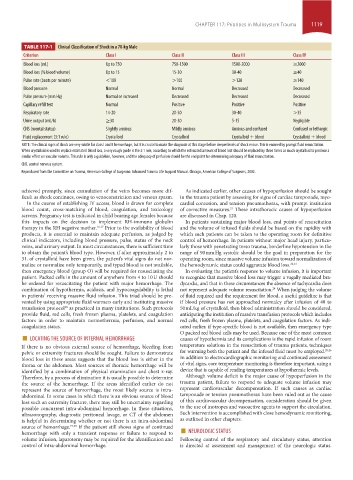
TABLE 117-1 Clinical Classification of Shock in a 70-kg Male
Criterion Class i Class ii Class iii Class iV
Blood loss (mL) Up to 750 750-1500 1500-2000 ≥2000
Blood loss (% blood volume) Up to 15 15-30 30-40 ≥40
Pulse rate (beats per minute) <100 >100 >120 ≥140
Blood pressure Normal Normal Decreased Decreased
Pulse pressure (mm Hg) Normal or increased Decreased Decreased Decreased
Capillary refill test Normal Positive Positive Positive
Respiratory rate 14-20 20-30 30-40 >35
Urine output (mL/h) ≥30 20-30 5-15 Negligible
CNS (mental status) Slightly anxious Mildly anxious Anxious and confused Confused or lethargic
Fluid replacement (3:1 rule) Crystalloid Crystalloid Crystalloid + blood Crystalloid + blood
NOTE: The clinical signs of shock are very subtle for class I and II hemorrhage, but it is crucial to make the diagnosis at this stage before deeper levels of shock ensue. This is ensured by prompt fluid resuscitation.
When crystalloid is used to replace estimated blood loss, a very rough guide is the 3:1 rule, according to which the estimated amount of blood lost should be replaced by three times as much crystalloid to produce a
similar effect on vascular volume. This rule is only a guideline, however, and the adequacy of perfusion should be the end point for determining adequacy of fluid resuscitation.
CNS, central nervous system.
Reproduced from the Committee on Trauma, American College of Surgeons: Advanced Trauma Life Support Manual. Chicago, American College of Surgeons, 2002.
achieved promptly, since cannulation of the veins becomes more dif- As indicated earlier, other causes of hypoperfusion should be sought
ficult as shock continues, owing to venoconstriction and venous spasm. in the trauma patient by assessing for signs of cardiac tamponade, myo-
In the course of establishing IV access, blood is drawn for complete cardial contusion, and tension pneumothorax, with prompt institution
blood count, cross-matching of blood, coagulation, and toxicology of corrective measures. These intrathoracic causes of hypoperfusion
21
screens. Pregnancy test is indicated in child bearing age females because are discussed in Chap. 120.
this impacts on the decision to implement RH-immuno globulin In patients sustaining major blood loss, end points of resuscitation
therapy in the RH negative mother. 16,17 Prior to the availability of blood and the volume of infused fluids should be based on the rapidity with
products, it is essential to maintain adequate perfusion, as judged by which such patients can be taken to the operating room for definitive
clinical indicators, including blood pressure, pulse, status of the neck control of hemorrhage. In patients without major head injury, particu-
veins, and urinary output. In most circumstances, there is sufficient time larly those with penetrating torso trauma, borderline hypotension in the
to obtain the patient’s blood type. However, if after approximately 2 to range of 90 mm Hg systolic should be the goal in preparation for the
3 L of crystalloid have been given, the patient’s vital signs do not nor- operating room, since massive volume infusion toward normalization of
malize or normalize only temporarily, and typed blood is not available, the hemodynamic status could aggravate blood loss. 22,23
then emergency blood (group O) will be required for resuscitating the In evaluating the patient’s response to volume infusion, it is important
patient. Packed cells in the amount of anywhere from 4 to 10 U should to recognize that massive blood loss may trigger a vagally mediated bra-
be ordered for resuscitating the patient with major hemorrhage. The dycardia, and that in these circumstances the absence of tachycardia does
combination of hypothermia, acidosis, and hypocoagulability is lethal not represent adequate volume resuscitation. When judging the volume
24
in patients’ receiving massive fluid infusion. This triad should be pre- of fluid required and the requirement for blood, a useful guideline is that
vented by using appropriate fluid warmers early and instituting massive if blood pressure has not approached normalcy after infusion of 40 to
transfusion protocol as practiced in many institutions. Such protocols 50 mL/kg of crystalloid, then blood administration should be considered,
18
provide fluid, red cells, fresh frozen plasma, platelets, and coagulation anticipating the institution of massive transfusion protocols which includes
factors in order to maintain normothermia, perfusion, and normal red cells, fresh frozen plasma, platelets, and coagulation factors. As indi-
coagulation status. cated earlier, if type-specific blood is not available, then emergency type
■ LOCATING THE SOURCE OF INTERNAL HEMORRHAGE O packed red blood cells may be used. Because one of the most common
causes of hypothermia and its complications is the rapid infusion of room
If there is no obvious external source of hemorrhage, bleeding from temperature solutions in the resuscitation of trauma patients, techniques
pelvic or extremity fractures should be sought. Failure to demonstrate for warming both the patient and the infused fluid must be employed. 25,26
blood loss in these areas suggests that the blood loss is either in the In addition to electrocardiographic monitoring and continued assessment
thorax or the abdomen. Most sources of thoracic hemorrhage will be of vital signs, core temperature monitoring is therefore important, using a
identified by a combination of physical examination and chest x-ray. device that is capable of reading temperatures at hypothermic levels.
Therefore, by a process of elimination it is usually possible to determine Although volume deficit is the major cause of hypoperfusion in the
the source of the hemorrhage. If the areas identified earlier do not trauma patient, failure to respond to adequate volume infusion may
represent the source of hemorrhage, the most likely source is intra- represent cardiovascular decompensation. If such causes as cardiac
abdominal. In some cases in which there is an obvious source of blood tamponade or tension pneumothorax have been ruled out as the cause
loss such an extremity fracture, there may still be uncertainty regarding of this cardiovascular decompensation, consideration should be given
possible concurrent intra-abdominal hemorrhage. In these situations, to the use of inotropes and vasoactive agents to support the circulation.
ultrasonography, diagnostic peritoneal lavage, or CT of the abdomen Such intervention is accomplished with close hemodynamic monitoring,
is helpful in determining whether or not there is an intra-abdominal as outlined in other chapters.
hemorrhage with only a transient response or failure to respond to ■ NEUROLOGIC STATUS
If the patient still shows signs of continued
source of hemorrhage.
19,20
volume infusion, laparotomy may be required for the identification and Following control of the respiratory and circulatory status, attention
control of intra-abdominal hemorrhage. is directed at assessment and management of the neurologic status.
section10.indd 1119 1/20/2015 9:20:10 AM