
Page 1605 - Hall et al (2015) Principles of Critical Care-McGraw-Hill
P. 1605
1124 PART 10: The Surgical Patient
related to motor vehicle collisions. Deficits in memory and learning are insignificant rate in humans and gene therapy is in the early research
8
5
common after TBI and may be related to frontal lesions. 6 phase without clinical application. The prevention of secondary injury
9
is the major goal in the treatment and optimization of outcomes of the
PRIMARY AND SECONDARY TBI patient after head injury. The optimal critical care of the head injured
patient requires the provision and maintenance of a homeostatic
A conceptual framework with which to care for neurologically injured environment that leads to recovery of potentially salvageable injured
patients is based on the classification of injury that occurs immediately or ischemic brain and prevents or mitigates insults that would render
after any insult, called primary injury, and all subsequent injuries, termed injured cells dead.
secondary injuries (Fig. 118-8). In TBI, primary injury occurs at the
moment of trauma and is the result of direct damage to brain tissue. After INITIAL STABILIZATION, IMAGING, AND MANAGEMENT
the primary injury, the remaining brain tissue consists of healthy tissue,
injured (or ischemic) tissue, and dead tissue. All subsequent brain injuries A moderate to severely head injured patient is a trauma patient with rapid
are termed secondary injuries and will result in further neuronal inju- triage and stabilization beginning in the field and transport, ideally to a neu-
ries and death over hours to days after the primary injury. Secondary rotrauma center (or the most appropriate hospital within range), facilitated
injuries may be caused by brain edema, hematoma expansion or delayed by emergency medical personnel, to continued evaluation and stabilization
hemorrhage, intracranial hypertension, herniation, hypotension, hypox- in the emergency department utilizing the ATLS protocol by trauma sur-
emia, hypercarbia or hypocarbia, circulatory or respiratory arrest, seizures, geons and neurosurgeons, followed by transport to radiology for diagnostic
vasospasm, and severe electrolyte disturbances. The pathophysiology of imaging or operating room for acute decompression of intracranial mass
secondary TBI involves impaired cerebrovascular autoregulation, cellular lesions, or ICU—the order of which is determined by the nature of the acute
metabolic dysfunction, and inadequate cerebral oxygenation. injuries. Head trauma associated with cervical spine injury and stabiliza-
On the cellular and molecular level, secondary injury results from tion of the spine (eg, cervical collar) is maintained until the spine is cleared
lactic acid production and depletion of ATP due to anaerobic glycolysis, (see Chap. 119, Spinal Injuries). Patients are admitted to the ICU depending
increased membrane permeability due to ion pump failure, and activa- on the risk or development of respiratory or circulatory failure, organ fail-
tion of voltage dependent calcium and sodium channels resulting in ure, and shock, and the severity of brain injury requiring close monitoring.
influx of calcium leading to activation of catabolic enzymes and free radi- After initial stabilization, patients routinely undergo computed tomog-
cal formation that leads to progressive damage to both intracellular and raphy (CT) imaging which provides immediate information regarding
nuclear structures leading to membrane failure, cytotoxic brain edema, the presence or absence of skull and spinal fractures, foreign objects,
necrosis, and apoptosis. The release of excitotoxic substances including contusions, extracranial and intracranial hemorrhages, edema, hydro-
7
the amino acids glutamate and aspartate damages adjacent neurons lead- cephalus, and herniation. Neurological examination may be limited
ing to further injury. Damage to the endothelial layer of the blood brain by depressed consciousness. Repeat or serial CT scans are useful to
barrier leads to vasogenic edema as well, but cytotoxic edema, which determine the etiology of acute deterioration or failure to improve and
7
does not respond to steroids, is more important after TBI. Primary as well to assess changes in initial lesions. When clinical deficits are not
as secondary injury results in the release of proinflammatory cytokines explained by CT findings, magnetic resonance imaging (MRI) is more
such as tumor necrosis factor, interleukin-1-β, and interleukin-6; pros- sensitive to assess the degree of DAI which is inferred by the presence of
taglandins, leukotrienes, and activation of complement and coagulation punctuate white matter hemorrhages, but such findings may be absent on
systems, neutrophils, macrophages, and lymphocytes that lead to further both CT and MRI. CT scanning is the primary study in patients with
10
endothelial damage and up regulation of cellular adhesion molecules penetrating head trauma associated with metallic foreign bodies where
such as P-selectin, intercellular adhesion molecules (ICAM-1), and vas- MRI scanning is contraindicated. Multidetector CT scanning (MDCT)
cular adhesion molecules (VCAM-1) that further facilitate the influx of allows three-dimensional imaging that may assist in preoperative
leukocytes into tissues leading to further secondary brain damage. 7 preparation. CSF leaks can present as CSF otorrhea or rhinorrhea and
10
Cellular necrosis occurs as the result of severe mechanical and ischemia- can be diagnosed with nuclear or CT cisternography, especially if the
hypoxia-induced injury. Apoptosis or programmed cell death may occur possible source is not clear on initial imaging. Cisternography performed
in cells that initially appear structurally intact and have adequate ATP after the injection of intrathecal contrast is more specific in identifying
and membrane potentials. Over hours to days after the injury, an imbal- the anatomical location of the leak, while nuclear cisternography is more
ance between pro- and antiapoptotic proteins with consecutive activa- sensitive to the existence of the leak but not its precise location.
tion and deactivation of caspases representing specific proteases of the Upon admission to the ICU, a tertiary head-to-toe examination is
interleukin-converting enzyme family are felt to be the most important performed to identify any potentially missed traumatic injuries and assess
11
mediators of apoptosis. 7 the current neurological exam noting any change that has occurred since
The prevention of primary head injury is a major public health the last described exam. The head is examined for ecchymoses, lacerations,
concern. Neurogenesis, the regeneration of neurons, occurs at an deformities, signs of basilar skull fracture (raccoon eyes, Battle sign), or
CSF leak (rhinorrhea or otorrhea). Neurological examination is focused on
assessment of the overall mental status, cranial nerves III and VI, pupillary
responses, oculocephalic (doll’s eyes), corneal and gag reflexes, deep tendon
Healthy reflexes, any a symmetry or focality of extremity movement, the presence
of pathologic reflexes (eg, Babinski sign or decorticate or decerebrate pos-
turing), and sensation. Bilateral and dilated fixed pupils indicate brain stem
Injured/Ischemic injury; unilateral or bilateral dilated fixed pupils may occur with cerebral
herniation, as does decerebrate posturing. Hypoxemia, shock, and hypo-
thermia may cause pupillary dilation and abnormal pupillary responses.
Dead Papilledema is usually not immediately seen in acute TBI with intracranial
hypertension (IH) and is a later finding. A negative neurologic exam does
12
not rule out significant intracranial injuries and intracranial hematomas
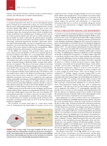
FIGURE 118-8. Conceptual model of brain injury. Immediately after TBI or any acute may be delayed, occurring days to weeks after the initial insult.
13
neurological insult, there are damaged (dead or injured) brain cells (red arrows) and uninjured or The Glasgow Coma Scale (GCS) (Fig. 118-9) can be used to deter-
“healthy” brain cells (primary injury). The central goal of care after TBI is the prevention of additional mine the severity of head injury, for serial assessment, and has prog-
brain cell injury or death (secondary injury) (red arrows) and the maintenance of an environment nostic implications. It is based on the best eye opening response, verbal
maximally conducive to recovery of the potentially salvageable injured cells (green arrow). response, and motor response. A score of ≤8 indicates severe TBI;
section10.indd 1124 1/20/2015 9:20:15 AM