
Page 1621 - Hall et al (2015) Principles of Critical Care-McGraw-Hill
P. 1621
1140 PART 10: The Surgical Patient
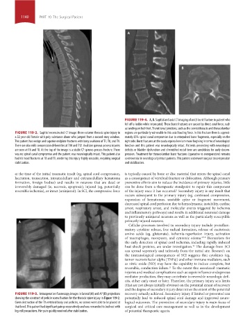
FIGURE 119-4. A, B. Sagittal and axial CT imaging of an L3 burst fracture in patient who
fell off a ladder while intoxicated. These burst fractures are caused by direct axial force, such
as landing on both feet. Transitional junctions, such as the cervicothoracic and thoracolumbar
FIGURE 119-2. Sagittal reconstructed CT image: three-column thoracic spine injury in regions are particularly vulnerable to this axial loading force. In this fracture there is approxi-
a 32 year-old female with poly substance abuse who jumped from a second story window. mately 85% spinal canal compromise due to retropulsed bone fragments, especially on the
This patient has wedge and superior endplate fractures with bony avulsions of T7, T8, and T9. right side. Burst fractures at the cauda equina level are more forgiving in terms of neurological
There are also mild compression deformities at T10 and T12. Avulsion spinous process injuries function and this patient was neurologically intact. Patients presenting with neurological
are seen at T6 and T8. At the top of the image is a stable C7 spinous process fracture. There deficits or bladder dysfunction and diminished rectal tone are candidates for early decom-
was no spinal canal compromise and the patient was neurologically intact. This patient also pression. Treatment for thoracolumbar burst fractures (operative vs nonoperative) remains
had rib head fractures at T8 and T9, rendering this injury highly unstable, requiring surgical controversial in neurologically intact patients. This patient underwent surgical decompression
stabilization. and stabilization.
at the time of the initial traumatic insult (eg, spinal cord compression, is typically caused by bone or disc material that enters the spinal canal
laceration, transection, intramedullary and extramedullary hematoma as a consequence of vertebral fracture or dislocation. Although primary
formation, foreign bodies) and results in neurons that are dead or preventive efforts aim to reduce the incidence of primary injuries, little
irreversibly damaged (ie, necrosis, apoptosis), injured (eg, potentially can be done from a therapeutic standpoint to repair this component
reversible ischemia), or intact (uninjured). In SCI, the compressive force of the injury once it has occurred. Secondary injury is any insult that
4
occurs subsequent to the primary injury (eg, continued compression,
expansion of hematomas, unstable spine or fragment movement,
decreased spinal cord perfusion due to hemodynamic instability, cardiac
arrest, respiratory arrest, and molecular events triggered by ischemia
and inflammatory pathways) and results in additional neuronal damage
to previously uninjured neurons as well as the particularly susceptible
primarily injured neurons.
Cellular processes involved in secondary injury include proinflam-
matory cytokine release, free radical formation, release of excitotoxic
amino acids (eg, glutamate), ischemia-reperfusion injury, activation
of macrophages, vasospasm, and cytotoxic edema. 4,7-9 Biomarkers for
the early detection of spinal cord ischemia, including rapidly induced
heat shock proteins, are under investigation. The damage from SCI
10
can spread superiorly and inferiorly from the initial site. Research on
the immunological consequences of SCI suggests that cytokines (eg,
tumor necrosis factor alpha [TNFa]) and other immune mediators, such
as nitric oxide (NO) may have the capability to induce complete, but
reversible, conduction failure. To the extent that associated traumatic
11
injuries and medical complications such as sepsis influence endogenous
mediator production, they may contribute to reversible neurologic defi-
cits, either early onset or later. Therefore, the primary injury sets limits
(that are not always initially obvious) on the potential extent of recovery
and the degree of secondary injury determines the extent of the potential
FIGURE 119-3. Intraoperative fluoroscopy images in lateral (A) and AP (B) projections recovery actually achieved. Secondary injury if limited or prevented can
showing the construct of pedicle screw fixation for the thoracic spine injury in Figure 119-2. potentially lead to reduced spinal cord damage and improved neuro-
Given destruction of the T8 vertebral body and pedicles, no screws were able to be placed at logical outcomes. The prevention of secondary injury is main focus of
that level. This patient had significant pain preoperatively and was remanded to bed rest with surgical and critical care management as well as in the development
log-roll precautions. Her pain quickly resolved after stabilization. of potential therapeutic agents.
section10.indd 1140 1/20/2015 9:20:27 AM