
Page 1622 - Hall et al (2015) Principles of Critical Care-McGraw-Hill
P. 1622
CHAPTER 119: Spinal Injuries 1141
GENERAL MANAGEMENT ISSUES AFTER ACUTE SCI detect only 60% to 80% of fractures; a significant number of fractures
are not visible, even when three views of the spine are obtained. In
1
Patients with acute SCI are managed in a multidisciplinary fashion from the cervical spine, MDCT detects 97% to 100% of fractures, provides
the accident scene by emergency response personnel, to triage and sta- finer anatomical delineation of the bony spinal canal, and also depicts
bilization in the emergency department by trauma or general surgeons epidural hemorrhages, significant soft tissue abnormalities such as trau-
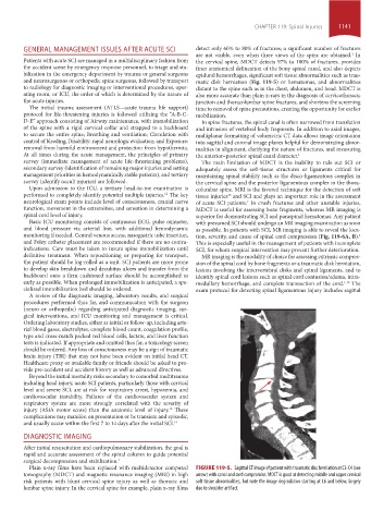
and neurosurgeons or orthopedic spine surgeons, followed by transport matic disk herniation (Fig. 119-5) or hematomas, and abnormalities
to radiology for diagnostic imaging or interventional procedures, oper- distant to the spine such as in the chest, abdomen, and head. MDCT is
ating room, or ICU, the order of which is determined by the nature of also more accurate than plain x-rays in the diagnosis of cervicothoracic
the acute injuries. junction and thoracolumbar spine fractures, and shortens the screening
The initial trauma assessment (ATLS—acute trauma life support) time to removal of spine precautions, creating the opportunity for earlier
protocol for life-threatening injuries is followed utilizing the “A-B-C- mobilization.
D-E” approach consisting of Airway maintenance, with immobilization In spine fractures, the spinal canal is often narrowed from translation
of the spine with a rigid cervical collar and strapped to a backboard and intrusion of vertebral body fragments. In addition to axial images,
to secure the entire spine; Breathing and ventilation; Circulation with multiplanar formatting of volumetric CT data allows image orientation
control of bleeding; Disability: rapid neurologic evaluation; and Exposure: into sagittal and coronal image planes helpful for demonstrating abnor-
removal from harmful environment and protection from hypothermia. malities in alignment, clarifying the nature of fractures, and measuring
At all times during the acute management, the principles of primary the anterior–posterior spinal canal diameter. 1
survey (immediate management of acute life-threatening problems), The main limitation of MDCT is the inability to rule out SCI or
secondary survey (identification of remaining major injuries and setting adequately assess the soft-tissue structures or ligaments critical for
management priorities in hemodynamically stable patients), and tertiary maintaining spinal stability such as the disco-ligamentous complex in
survey (identify occult injuries) are followed. the cervical spine and the posterior ligamentous complex in the thora-
Upon admission to the ICU, a tertiary head-to-toe examination is columbar spine. MRI is the favored technique for the detection of soft
performed to completely identify potential multiple injuries. The key tissue injuries and SCI and plays an important role in the assessment
12
15
neurological exam points include level of consciousness, cranial nerve of acute SCI patients. In crush fractures and other unstable injuries,
1
function, movement in the extremities, and sensation in determining a MDCT is useful for assessing bone fragments, whereas MR imaging is
spinal cord level of injury. superior for demonstrating SCI and paraspinal hematomas. Any patient
Basic ICU monitoring consists of continuous ECG, pulse oximeter, with presumed SCI should undergo an MR imaging examination as soon
and blood pressure via arterial line, with additional hemodynamic as possible. In patients with SCI, MR imaging is able to reveal the loca-
monitoring if needed. Central venous access, nasogastric tube insertion, tion, severity, and cause of spinal cord compression (Fig. 119-6A, B).
1
and Foley catheter placement are recommended if there are no contra- This is especially useful in the management of patients with incomplete
indications. Care must be taken to insure spine immobilization until SCI, for whom surgical intervention may prevent further deterioration.
definitive treatment. When repositioning or preparing for transport, MR imaging is the modality of choice for assessing extrinsic compres-
the patient should be log-rolled as a unit. SCI patients are more prone sion of the spinal cord by bone fragments or a traumatic disk herniation,
to develop skin breakdown and decubitus ulcers and transfer from the lesions involving the intervertebral disks and spinal ligaments, and to
backboard onto a firm cushioned surface should be accomplished as identify spinal cord lesions such as spinal cord contusion/edema, intra-
early as possible. When prolonged immobilization is anticipated, a spe- medullary hemorrhage, and complete transsection of the cord. The
1,16
cialized immobilization bed should be ordered. exam protocol for detecting spinal ligamentous injury includes sagittal
A review of the diagnostic imaging, laboratory results, and surgical
procedures performed thus far, and communication with the surgeon
(neuro or orthopedic) regarding anticipated diagnostic imaging, sur-
gical interventions, and ICU monitoring and management is critical.
Ordering laboratory studies, either as initial or follow-up, including arte-
rial blood gases, electrolytes, complete blood count, coagulation profile,
type and cross-match packed red blood cells, lactate, and liver function
tests is indicated. If appropriate and omitted thus far, a toxicology screen
should be ordered. Any loss of consciousness may be a sign of traumatic
brain injury (TBI) that may not have been evident on initial head CT.
Healthcare proxy or available family or friends should be asked to pro-
vide pre-accident and accident history as well as advanced directives.
Beyond the initial mortality risks secondary to comorbid multitrauma
including head injury, acute SCI patients, particularly those with cervical
level and severe SCI, are at risk for respiratory arrest, hypoxemia, and
cardiovascular instability. Failures of the cardiovascular system and
respiratory system are more strongly correlated with the severity of
injury (ASIA motor score) than the anatomic level of injury. These
13
complications may manifest on presentation or be transient and episodic,
and usually occur within the first 7 to 14 days after the initial SCI. 14
DIAGNOSTIC IMAGING
After initial resuscitation and cardiopulmonary stabilization, the goal is
rapid and accurate assessment of the spinal column to guide potential
surgical decompression and stabilization. 1
Plain x-ray films have been replaced with multidetector computed FIGURE 119-5. Sagittal CT image of patient with traumatic disc herniation at C3-C4 (see
tomography (MDCT) and magnetic resonance imaging (MRI) in high arrow) with canal and cord compromise. MDCT is good at detecting middle and upper cervical
risk patients with blunt cervical spine injury as well as thoracic and soft tissue abnormalities, but note the image degradation starting at C6 and below, largely
lumbar spine injury. In the cervical spine for example, plain x-ray films due to shoulder artifact.
section10.indd 1141 1/20/2015 9:20:28 AM