
Page 1835 - Hall et al (2015) Principles of Critical Care-McGraw-Hill
P. 1835
1304 PART 11: Special Problems in Critical Care
■
TABLE 129-21 Histologic Findings for Deep Fungal Infections PRESSURE ULCERS
Condition Staining Technique Key Histologic Findings The incidence of pressure ulcers in the ICU varies widely in the litera-
ture. Continuous pressure over a bony site obstructs microcirculation,
Blastomycosis KOH mount Round, refractile spherical cells leading to tissue ischemia and necrosis. ICU patients have multiple risk
with broad-based budding factors for developing pressure ulcers, which can occur in as little as
Histoplasmosis H & E stain, Giemsa stain Small yeast-like spores within 2 hours under certain conditions. Mechanical ventilation, limited mobil-
macrophages ity, hypoperfusion, and the use of vasoactive drugs may increase the risk
Cryptococcus India ink stain Round to ovoid spores with of pressure ulcers. The development of pressure ulcers leads to increased
large capsules mortality rates, costs, and lengths of hospital stays.
A National Pressure Ulcer Advisory Panel in 1998 developed the most
Aspergillosis H & E stain, PAS, silver methenamine stain Branching septate hyphae widely used staging system of pressure ulcers. It encompasses four grades.
129
Mucormycosis H & E stain, PAS, silver methenamine Large, long, nonseptate Stage I: A stage I pressure ulcer is an observable pressure-related
stain hyphae that may invade alteration of intact skin that may display a different skin temperature
vascular structures
(warmth or coolness), tissue consistency (firm or boggy feel), or sen-
sation (pain, itching). The ulcer appears as a defined area of persistent
redness in lightly pigmented skin, but can appear with red, blue, or
findings are outlined in Table 129-21. Systemic treatment with itracon- purple hues in darker skin tones.
azole, fluconazole, amphotericin B, or caspofungin is required to control
each of these diseases, as is extensive debridement of all necrotic tissue. 128 Stage II: A stage II ulcer is defined as partial-thickness skin loss
involving epidermis, dermis, or both. The ulcer is superficial and
SELECTED DERMATOSES presents clinically as an abrasion, blister, or shallow crater.
■ MILIARIA Stage III: The stage III ulcer is characterized by full-thickness skin loss
involving damage to, or necrosis of, subcutaneous tissue that may extend
Miliaria, sometimes referred to as heat rash, is caused by obstruction down to, but not through, underlying fascia. The ulcer presents clinically
of the eccrine (sweat) ducts at a variety of levels causing sweat reten- as a deep crater with or without undermining of adjacent tissue.
tion. Miliaria can be classified into three groups, based on the levels Stage IV: This ulcer is characterized by full-thickness skin loss with
of ductal obstruction: miliaria crystallina, miliaria rubra, and miliaria extensive destruction, tissue necrosis, or damage to muscle, bone, or
profunda. 128 supporting structures (eg, tendon, joint capsule).
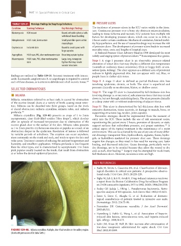
Miliaria crystallina (Fig. 129-40) presents as crops of 1 to 2 mm Preventive strategies should be implemented from the moment of
asymptomatic, clear fluid-filled vesicles (“dew drops”), which develop entry into the ICU. These include the use of risk assessment scales,
after an episode of increased temperature due to obstruction of the repositioning the patient every 2 hours, provision of dynamic or static
eccrine gland close to the surface of the skin. Miliaria rubra presents support surfaces that redistribute pressure, and proper nutrition. A
130
as 1 to 2 mm pruritic erythematous macules or papules as a result of an critical aspect of the topical treatment is the maintenance of a moist
obstruction deeper in the epidermis. Resolution of lesions is followed environment. This can be achieved by the use of any one of many differ-
by variable periods of anhidrosis. The eruption can occur anywhere, ent dressings: transparent films, hydrocolloids, alginates, foams, hydro-
most commonly on the trunk and neck, and tends to spare the face and gels, or hydrofibers marketed for pressure ulcer care. These dressings
volar areas. Treatment consists of reducing the ambient temperature and require few changes, so they result in less need for nursing care, faster
humidity, and emollient application. Miliaria profunda is less frequent healing, and decreased infection. Gauze dressings, particularly wet to
than the other types, and is characterized by asymptomatic 1 to 3 mm dry dressings, are to be avoided because they allow the wound to dry
pink papules usually located on the trunk, that result from obstruction and, as such, slow healing. Surgery may be attempted for recalcitrant,
131
at or below the dermal-epidermal junction. full-thickness ulcers. However, recurrence rates are high.
KEY REFERENCES
• Badia M, Servia L, Casanova JM, et al. Classification of dermato-
logical disorders in critical care patients: A prospective observa-
tional study. J Crit Care. 2013; 28:220-228.
• Bigby M, Jick S, Jick H, Arndt K. Drug-induced cutaneous reactions:
a report from the Boston Collaborative Drug Surveillance Program
on 15438 consecutive inpatients, 1975 to 1982. JAMA. 1986;256:3358.
• Bodey GP, Jadeja L, Elting L. Pseudomonas bacteremia. Retro-
spective analysis of 410 episodes. Arch Intern Med. 1985;145:1621.
• Emre S, Emre C, Akoglu G, et al. Evaluation of dermato-
logical consultations of patients treated in intensive care units.
Dermatology. 2013; 226:75-80.
• Fiorentino DF. Cutaneous vasculitis. J Am Acad Dermatol.
2003;48:311.
• Harenberg J, Huhle G, Wang L, et al. Association of heparin-
induced skin lesions, intracutaneous tests, and heparin-induced
IgG. Allergy. 1999;54:473.
• Kahn JM, Kress JP, Hall JB. Skin necrosis after extravasation of
low-dose vasopressin administered for septic shock. Crit Care
FIGURE 129-40. Miliaria crystallina. Multiple, clear-fluid vesicles on the axillary region. Med. 2002;30:1899.
(Used with permission of Dr Aisha Sethi.)
section11.indd 1304 1/19/2015 10:56:00 AM