
Page 1833 - Hall et al (2015) Principles of Critical Care-McGraw-Hill
P. 1833
1302 PART 11: Special Problems in Critical Care
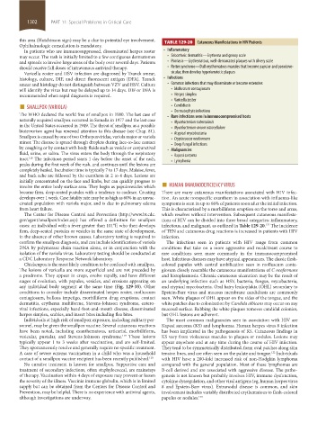
this area (Hutchinson sign) may be a clue to potential eye involvement. TABLE 129-20 Cutaneous Manifestations in HIV Patients
Ophthalmologic consultation is mandatory.
In patients who are immunosuppressed, disseminated herpes zoster • Inflammatory
may occur. The rash is initially limited to a few contiguous dermatomes • Seborrheic dermatitis—Erythema and greasy scale
and spreads to involve large areas of the body over several days. Patients • Psoriasis—Erythematous, well-demarcated plaques with silvery scale
should receive full doses of intravenous antiviral therapy. • Reiter syndrome—Dull erythematous macules that become papular and pseudove-
Varicella zoster and HSV infection are diagnosed by Tzanck smear, sicular, then develop hyperkeratotic plaques
histology, culture, DIF, and direct fluorescent antigen (DFA). Tzanck • Infections
smear and histology do not distinguish between VZV and HSV. Culture • Common infections that may disseminate or become extensive:
will identify the virus but may be delayed up to 14 days. DIF or DFA is • Molluscum contagiosum
recommended when rapid diagnosis is required. • Herpes simplex
■ SMALLPOX (VARIOLA) • Varicella zoster
• Candidiasis
• Dermatophyte infections
The WHO declared the world free of smallpox in 1980. The last case of • Rare infections seen in immunocompromised hosts
naturally acquired smallpox occurred in Somalia in 1977 and the last case • Mycobacterium tuberculosis
in the United States occurred in 1949. The threat of smallpox as a possible • Mycobacterium avium intracellulare
bioterrorism agent has renewed attention to this disease (see Chap. 81). • Atypical mycobacteria
Smallpox is caused by one of two Orthopoxviridae, variola major or variola • Cryptococcus neoformans
minor. The disease is spread through droplets during face-to-face contact • Deep fungal infections
by coughing or by contact with body fluids such as vesicle or conjunctival • Malignancies
fluid, urine, or saliva. The virus enters the body through the respiratory • Kaposi sarcoma
tract. The infectious period starts 1 day before the onset of the rash, • Lymphoma
118
peaks during the first week of the rash, and continues until the lesions are
completely healed. Incubation time is typically 7 to 17 days. Malaise, fever,
and back ache are followed by the exanthem in 2 to 4 days. Lesions are
involve the entire body surface area. They begin as papulovesicles which ■ HUMAN IMMUNODEFICIENCY VIRUS
initially concentrated on the face and limbs, but can quickly progress to
become firm, deep-seated pustules with a tendency to coalesce. Crusting There are many cutaneous manifestations associated with HIV infec-
develops over 1 week. Case fatality rate may be as high as 60% in an unvac- tion. An acute nonspecific exanthem in association with influenza-like
cinated population with variola major, and is due to pulmonary edema symptoms is seen in up to 66% of patients soon after the initial infection.
from heart failure. This is characterized by a morbilliform eruption on the torso and arms,
The Center for Disease Control and Prevention (http://www.bt.cdc. which resolves without intervention. Subsequent cutaneous manifesta-
gov/agent/smallpox/index.asp) has offered a definition for smallpox tions of HIV can be divided into three broad categories: inflammatory,
cases: an individual with a fever greater than 101°F, who then develops infectious, and malignant, as outlined in Table 129-20. The incidence
121
firm, deep-seated pustules or vesicles in the same state of development, of TEN and cutaneous drug reactions is increased in patients with HIV
in the absence of other known causes. Laboratory testing is required to infection.
confirm the smallpox diagnosis, and can include identification of variola The infections seen in patients with HIV range from common
DNA by polymerase chain reaction alone, or in conjunction with the conditions that take on a more aggressive and recalcitrant course to
isolation of the variola virus. Laboratory testing should be conducted at rare conditions seen more commonly in the immunocompromised
a CDC Laboratory Response Network laboratory. host. Infectious diseases may have atypical appearances. The classic flesh-
Chickenpox is the most likely condition to be confused with smallpox. colored papules with central umbilication seen in molluscum conta-
The lesions of varicella are more superficial and are not preceded by giosum closely resemble the cutaneous manifestations of C neoformans
a prodrome. They appear in crops, evolve rapidly, and have different and histoplasmosis. Chronic cutaneous ulceration may be the result of
stages of evolution, with papules, vesicles, and erosions appearing on an underlying infection such as HSV, bacteria, fungus, mycobacteria,
any individual body segment at the same time (Fig. 129-35). Other and atypical mycobacteria. Oral hairy leukoplakia (OHL) secondary to
conditions to consider include disseminated herpes zoster, molluscum Epstein-Barr virus and mucous membrane candidiasis are commonly
contagiosum, bullous impetigo, morbilliform drug eruptions, contact seen. White plaques of OHL appear on the sides of the tongue, and the
dermatitis, erythema multiforme, Stevens-Johnson syndrome, entero- white patches due to colonization by Candida albicans may occur on any
viral infections, especially hand-foot-and-mouth disease, disseminated mucosal surface. Rubbing the white plaques removes candidal colonies,
herpes simplex, scabies, and insect bites including flea bites. but OHL lesions are adherent.
Individuals at high risk of smallpox exposure, including military per- The most common malignancies seen in association with HIV are
sonnel, may be given the smallpox vaccine. Several cutaneous reactions Kaposi sarcoma (KS) and lymphomas. Human herpes virus 8 infection
have been noted, including exanthematous, urticarial, morbilliform, has been implicated in the pathogenesis of KS. Cutaneous findings in
vesicular, pustular, and Stevens-Johnson syndrome. These lesions KS vary from violaceous macules to plaques or nodules. Lesions may
119
typically appear 1 to 3 weeks after vaccination, and are self-limited. appear anywhere and at any time during the course of HIV infection.
They spontaneously resolve and generally require no specific treatment. They tend to be symmetrically distributed, form oval patches along skin
A case of severe eczema vaccinatum in a child who was a household tension lines, and are often seen on the palate and tongue. Individuals
122
contact of a smallpox vaccine recipient has been recently published. 120 with HIV have a 200-fold increased risk of non-Hodgkin lymphoma
No curative treatment is known for smallpox. Supportive care and compared with the general population. Most of these lymphomas are
treatment of secondary infections, often staphylococcal, are mainstays B-cell derived and are associated with aggressive disease. The patho-
of therapy. Vaccination within 4 days of exposure may prevent or lessen genesis is not known but probably involves HIV, immune dysfunction,
the severity of the illness. Vaccinia immune globulin, which is in limited cytokine dysregulation, and other viral antigens (eg, human herpes virus
supply but can be obtained from the Centers for Disease Control and 8 and Epstein-Barr virus). Extranodal disease is common, and skin
Prevention, may be helpful. There is no experience with antiviral agents, involvement includes variably distributed erythematous to flesh-colored
although investigations are underway. papules or nodules. 123
section11.indd 1302 1/19/2015 10:55:54 AM