
Page 1844 - Hall et al (2015) Principles of Critical Care-McGraw-Hill
P. 1844
CHAPTER 131: Hypothermia 1313
Clinical manifestations vary with the etiology of hypothermia, rapidity Arterial Blood Gases: Typically, pH values increase as body temperature
of cooling and the duration and severity of hypothermia. The severity of decreases (0.0147 increase for each 1°C decrease). Arterial pressures
hypothermia is classified based on core temperature as mild (35-32.2°C, of O 2 and CO 2 also decrease with a decrease in temperature (7.2% and
95-90°F), moderate (<32.2-28°C, <90-82°F), and severe (<28°C, 4.4%, respectively, for each 1°C decrease in temperature), and the oxy-
<82°F). 2,3,7,9,13,14 The classification for patients with traumatic injuries is hemoglobin dissociation curve is shifted to the left. However, because
more conservative due to worse outcomes, with a core temperature of all arterial blood gas samples are warmed to 37°C (98.6°F) before values
<32°C (90°F) considered severe hypothermia. 15,16 This classification has are measured, simply comparing uncorrected values measured at 37°C
implications for management because appropriate treatment depends on with the normal reference values at 37°C yields an accurate interpreta-
severity of the disorder, as described below. The onset of hypothermia tion. Respiratory acidosis and metabolic acidosis are common findings
is often insidious. Initial symptoms may be vague and include hunger, in patients with moderate and severe hypothermia. 20
nausea, dizziness, chills, pruritus, or dyspnea. Extremity stiffness, weak-
ness, and shivering may also be prominent. As core body temperature Complete Blood Count: An increase in hematocrit secondary to decrease
decreases, many patients no longer complain of cold, shivering disap- in plasma volume is common (2% for each 1°C decrease in temperature).
pears at temperatures below 32°C (<90°F), and muscles become rigid. A low initial hematocrit suggests bleeding or preexisting anemia. White
7,17
2,7
At this point, the level of consciousness becomes markedly altered and blood cell and platelet counts may decrease as temperature decreases.
systemic manifestations are readily evident. A severely hypothermic A normal or low white blood cell count cannot be used as an indicator
victim has a markedly decreased metabolic rate. As a consequence, the for the absence of infection.
cerebral ischemic tolerance during cardiocirculatory arrest is consider- Coagulation Profile: A physiologic coagulopathy occurs with hypothermia
ably longer in contrast with the normothermic state. 7,18,19 Therefore, one due to inhibition of coagulation factors. Hypothermia is associated with
has to be very careful in assessing brain death while a patient remains thrombocytopenia secondary to bone marrow suppression and splenic
hypothermic. Low temperatures cause the myocardium to become irrita- and hepatic sequestration, as well as reduction in platelet function.
ble and cardiovascular abnormalities are common. These may include Disseminated intravascular coagulation may also occur with rewarming.
18
initial tachycardia followed by progressive bradycardia with an increase Prolonged bleeding and clotting times are common. Prothrombin time
in systemic vascular resistance. Arrhythmias are common at core tem- and partial thromboplastin time may initially appear normal despite the
peratures below 32°C (90°F), and ventricular fibrillation may occur presence of clinical coagulopathy because the tests are performed after
spontaneously when the temperature is below 28°C (82°F). Systemic warming the blood sample to 37°C.
17
blood pressure is often decreased in patients with severe hypothermia.
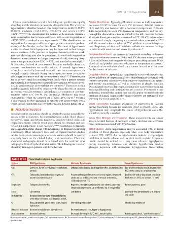
Other clinical manifestations of hypothermia are listed in Table 131-2. Serum Electrolytes: Recurrent evaluation of electrolytes is essential
■ LABORATORY EVALUATION during rewarming because no consistent effect is present. Hypo- and
hyperkalemia may complicate the course of hypothermia and either
Initial laboratory evaluations should be obtained to assess metabolic sta- should be promptly corrected.
tus and organ dysfunction. Recommended tests include blood glucose, Serum Urea Nitrogen and Creatinine: These measurements are almost
electrolytes, renal and hepatic functions, complete blood count, and always elevated because of decreased urinary clearance and decreased
coagulation profile. Arterial blood gases should be obtained, and cor- renal perfusion associated with hypovolemia.
rection for temperature is not necessary. 7,20,21 Electrolytes, hematocrit,
and coagulation status change with rewarming, so frequent monitoring Blood Glucose: Acute hypothermia may be associated with an initial
is necessary. Other laboratory tests such as thyroid function studies, elevation of blood glucose, especially when core body temperature
cardiac isoenzymes, toxicologic screen, and cultures should be ordered is above 30°C (86°F), due to catecholamine-induced glycogenolysis,
selectively based on the clinical history and examination. Chest and inhibition of insulin release, and impaired insulin uptake. Exogenous
abdominal radiographs should be obtained with the need for other insulin should be avoided as it may cause rebound hypoglycemia
radiographs dictated by the clinical situation. The following are common during rewarming. Subacute and chronic hypothermia produce
laboratory findings in patients with hypothermia. glycogen depletion with subsequent hypoglycemia. Nevertheless,
TABLE 131-2 Clinical Manifestations of Hypothermia
System Mild Hypothermia Moderate Hypothermia Severe Hypothermia
CNS Confusion, slurred speech, impaired judgment, Lethargy, hallucinations, loss of pupillary reflex, EEG abnormalities Loss of cerebrovascular regulation, decline in
amnesia EEG activity, coma, loss of ocular reflex
CVS Tachycardia, increased cardiac output and Progressive bradycardia (unresponsive to atropine), decreased Decline in BP and cardiac output, ventricular
systemic vascular resistance cardiac output and BP, atrial and ventricular arrhythmias, fibrillation (<28°C) and asystole (<20°C)
J (Osborn) wave on ECG
Respiratory Tachypnea, bronchorrhea Hypoventilation (decreased rate and tidal volume), decreased Pulmonary edema, apnea
oxygen consumption and CO 2 production, loss of cough reflex
Renal Cold diuresis Cold diuresis Decreased renal perfusion and GFR, oliguria
Hematologic Increased hematocrit and decreased platelet, and anuria
white blood cell count, coagulopathy, and DIC
GI Ileus, pancreatitis, gastric stress ulcers, hepatic Altered drug metabolism Altered drug metabolism
dysfunction
Metabolic endocrine Increased metabolic rate, hyperglycemia Decreased metabolic rate, hyper- or hypoglycemia
Musculoskeletal Increased shivering Decreased shivering (<32°C, 90°F), muscle rigidity Patient appears dead, “pseudo-rigor mortis”
BP, blood pressure; CNS, central nervous system; CVS, cardiovascular system; DIC, disseminated intravascular coagulation; ECG, electrocardiogram; EEG, electroencephalogram; GFR, glomerular filtration rate; GI,
gastrointestinal.
section11.indd 1313 1/19/2015 10:56:05 AM