
Page 1845 - Hall et al (2015) Principles of Critical Care-McGraw-Hill
P. 1845
1314 PART 11: Special Problems in Critical Care
V4
V5
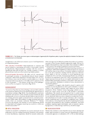
FIGURE 131-1. The J (Osborn) wave (arrows) appears on electrocardiograms of approximately 80% of hypothermic patients. In general, the amplitude and duration of the Osborn wave
are inversely related to core temperature.
hypoglycemia is one of the most common causes of mild hypothermia Other vital signs may be difficult to evaluate, but evidence of a spontane-
in a hospitalized patient. ous pulse or blood pressure should be aggressively sought. The use of a
Doppler ultrasound device may be necessary. Pulse oximetry is unlikely
Other Laboratory Abnormalities: Hyperamylasemia is common and to be accurate in the setting of hypothermia and poor perfusion.
may be related to a preexisting pancreatitis or pancreatitis induced Supplemental oxygen should be administered pending assessment of
by hypothermia. Hyperamylasemia correlates with the severity of oxygenation. Endotracheal intubation is indicated unless the patient is
hypothermia and with mortality rate. Variable elevation in creatine alert and has intact airway reflexes. Orotracheal intubation is preferred
phosphokinase levels may reflect underlying rhabdomyolysis.
due to the risk of traumatic bleeding with the nasal route. However,
Electrocardiographic Abnormalities: PR, QRS, and QT intervals may muscle rigidity of the jaw in moderate to severe hypothermia may
be prolonged secondary to hypothermia-induced slowed impulse preclude use of the oral route. Neuromuscular blockers are unlikely to
conduction. When body temperature decreases below 33°C (91.4°F), be effective at temperatures below 30°C (86°F) and should be avoided.
the J (Osborn) wave may be noted as a positive deflection in the left Topical vasoconstrictors and the use of a smaller endotracheal tube may
ventricular leads at the junction of the QRS and ST segments in 25% facilitate blind nasotracheal intubation in the patient with some spon-
to 30% of patients. 2,22-24 The presence of this wave is not pathogno- taneous respirations. Intubation is unlikely to induce dysrhythmias in
monic and has no prognostic implication (Fig. 131-1). hypothermic patients. 10
A nasogastric or orogastric tube should be placed in patients with
MANAGEMENT moderate or severe hypothermia to relieve gastric distention. A urinary
catheter is also essential to monitor urine output and assess volume
The severity of hypothermia, clinical findings, and comorbid conditions resuscitation efforts. Peripheral venous large gauge catheters are pre-
of the patient are important factors for determining the aggressiveness of ferred over central venous access due to the potential to precipitate
resuscitation techniques (Fig. 131-2). Once hypothermia is confirmed, dysrhythmias. The femoral vein is the preferred site if a central venous
assessment and treatment of the critically ill patient should take place catheter is needed. Intraarterial catheters for pressure monitoring should
simultaneously. Actions in all patients should include prompt removal of be used selectively. Pulmonary artery catheters are avoided due to poten-
wet clothing, protection against continued heat loss, continuous moni- tial dysrhythmias and the risk of vascular perforation. After rewarming,
toring of cardiac status, and avoidance of rough movement and excess invasive monitoring may be warranted in complicated cases. Patients
activity, which can precipitate ventricular fibrillation. 21,25-27 Patients with moderate or severe hypothermia should be handled gently because
received in the hospital with moderate or severe hypothermia should movement and manipulation may precipitate arrhythmias. Associated
be resuscitated until adequate rewarming has occurred or efforts are conditions requiring urgent intervention, such as traumatic injuries,
deemed unsuccessful. 14,25,26 hypoglycemia, or endocrinologic insufficiency, should be sought.
■ INITIAL STABILIZATION ■ VOLUME RESUSCITATION
Hypothermia should be confirmed by an accurate assessment of core Patients with moderate or severe hypothermia are volume depleted and
temperature with a low reading thermometer. In patients with moderate require rapid assessment of volume status and administration of fluids.
to severe hypothermia, temperature should be assessed continuously. A reasonable approach is to administer a 250- to 500-mL fluid challenge
section11.indd 1314 1/19/2015 10:56:06 AM