
Page 374 - Clinical Hematology_ Theory _ Procedures ( PDFDrive )
P. 374
358 PART 5 ■ Nonmalignant Leukocyte Disorders
800
Henke et al.
700 Health et al.
Davidson
n
o 600
i
t
a
l
u
p
o 500
p
0
0
0
,
0 400
0
1
r
e
p 300
s
e
s
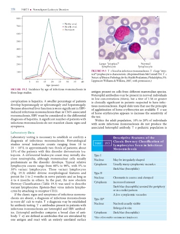
Ca 200 Large "atypical" Normal
lymphocyte lymphocyte
FIGURE 19.3 T m . T “ -
100
” m . (R m M C H. Te
Nature o Disease Pathology or the Health Pro essions, P , PA:
L W m & W k , , m .)
5 10 15 20 25 30 35 40
Age (years )
FIGURE 19.2 I m
. m m mm .
H m m
( ), :
m . A m -
m m m . m . R
B m m EBV- . T
m CMV-
m , EBV m .
. A m W , % %
m m m
m m . . T
Laboratory Data
L m Descriptive Features of the
m . H m Classic Dow ney Classi cation of
k m TABLE 19.3 Lymphocytes Seen in Infectious
× /L x m ; Mononucleosis
% m leu-
kopenia. A k m - Type I
, m Nucleus May be irregularly shaped
m .
m m % %, % Cytoplasm Usually many cytoplasmic vacuoles
% m . m Dark blue (basophilic)
(F . . ) x m Type II
m m Nucleus Chromatin is coarse and clumped
m . I ,
D C ( . ) Cytoplasm Increased amount
m . E -B m - Dark blue (basophilic) around the periphery
CD . or in a radial pattern
I m m m - A few cytoplasmic vacuoles
, m Type III*
m f m k . T m Nucleus Nucleoli usually visible
. T
m EBV - Enlarged in size
. H m - Cytoplasm Dark blue (basophilic)
. T m *This cell resembles an immature lymphocyte.