
Page 608 - Clinical Hematology_ Theory _ Procedures ( PDFDrive )
P. 608
592 PART 8 ■ Fundamentals of Hematological Analysis
Pleural Fluid
NOTE: This is a good time to complete Review Questions
related to preceding content. Anatomy of the Pleura
Te lungs lie in the thoracic (chest) cavity, where they are sepa-
rated by the heart in the mediastinum. Each lung is covered by
PLEURAL, PERITONEAL, AND a serous membrane, the visceral pleura (Fig. 29.4). T e interior
PERICARDIAL FLUIDS o the chest wall, the superior sur ace o the diaphragm, and the
lateral portion o the mediastinum are also lined by a thin mem-
Effusions: Transudates and Exudates brane, the parietal pleura. T e layers o the visceral and parietal
An effusion is an abnormal accumulation o f uid in a par- pleurae are contiguous, and the potential space between them
ticular cavity o the body. E usions in the pleural, pericar- on each side o the thorax orms the pleural cavity. However, the
dial, and peritoneal cavities are divided into transudates pleural cavity is not a true cavity. It becomes a cavity i an abnor-
and exudates. ransudates generally indicate that f uid has mal condition creates an excess accumulation o f uid or air in it.
accumulated because o the presence o a systemic disease. T e pleural cavity is lined by a single-cell layer o meso-
In contrast, exudates are usually associated with disorders thelial cells that orm the mesothelium. Mesothelial cells
such as inf ammation, in ection, and malignant conditions are supported by layers o connective tissue that contain an
involving the cells that line the sur aces o organs (e.g., lung extensive network o lymphatic vessels and blood capillar-
or abdominal organs). ies. Although the unction o the pleural space is obscure,
ransudates and exudates requently di er in character- the stretchable mesothelial cells that line this potential space
istics such as color and clarity and in total leukocyte cell provide the lungs and other intrathoracic organs with the
count. Classically, transudates have been considered to di er fexibility to expand and retract.
rom exudates based on the properties o speci c gravity and Pleural f uid is normally produced by the parietal pleura
total protein. T ese characteristics, however, are unreliable and absorbed by the visceral pleura as a continuous process.
in consistently di erentiating the two categories o e usions. Although healthy individuals rom 600 to 800 mL o f uid
For example, the mean values o total protein display consid- daily, the normal volume o f uid in each pleural space is esti-
erable overlap between transudates and exudates. mated at less than 10 mL. T is f uid is ormed by the ltration
A variety o physical and chemical properties need to be o blood plasma through the capillary endothelium. T e f uid
considered when f uids are categorized as transudates or is reabsorbed by lymphatic vessels and venules in the pleura.
exudates ( able 29.5). ransport in and out o the pleural space is dependent on the
balance o hydrostatic pressure in the capillary network o
the parietal and visceral pleurae and capillary permeability,
plasma oncotic pressure, and lymphatic reabsorption.
Comparison of Transudates
TABLE 29.5
and Exudates *
Characteristics Transudate Exudate
Physical Characteristics
pH 7.4–7.5 7.35–7.45
Speci c gravity <1.016 >1.016
Cellular Characteristics
Erythrocytes Few Variable
Leukocytes <1,000 >1,000
Chemical Analyses
Glucose level Equal to serum Possibly
decreased
Protein level <3.0 g/dL >3.0 g/dL
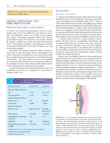
Pleural uid–serum ratio <0.5 >0.5 FIGURE 29.4 T oracocentesis. Sometimes it is necessary to insert
of protein a hypodermic needle through an intercostal space into the pleu-
LDH level <200 >200 IU/L ral cavity—the potential space between the parietal pleura lining
Pleural uid–serum ratio <2:3 (<0.6) >2:3 (>0.6) the pulmonary cavity and the visceral pleura covering the lung—to
of LDH † obtain a sample o pleural f uid or to remove blood or pus. o avoid
damage to the intercostal nerve and vessels, the needle is inserted
* Variations can be observed in examples of various conditions. superior to the rib, high enough to avoid the collateral branches.
† If nonhemolyzed, nonbloody effusion. (Reprinted rom Moore KL, Agur A. Essential Clinical Anatomy,
2nd ed, Philadelphia, PA: Lippincott Williams & Wilkins, 2002,
LDH, lactic dehydrogenase.
with permission.)