Page 613 - Clinical Hematology_ Theory _ Procedures ( PDFDrive )
P. 613

CHAPTER 29  ■  Body Fluid Analysis                        597


                                                                                                           Diaphragm


                                                                                                                                              Aorta


                                                                                                                                                             Superior recess

                                                              Porta hepatis                                                                                  of lesser sac


                                                          Lesser omentum
                                                                                                                                                          Celiac artery

                                                                                                                                                          Pancreas

                                                                                                                                                      Lesser sac
                                                                 Stomach
                                                                                                                                                 Superior mesenteric artery
                                                 Transverse mesocolon

                                                                                                                                                 Third part of duodenum
                                                        Transverse colon

                                                                                                                                                    Mesentery

                                                            Umbilicus
                                                                                                                                                       Greater sac


                                                                   Jejunum


                                                Inferior recess of lesser
                                                                             sac
                                                           Greater omentum
                                                                                                                                                                          Rectum


                                                                                                                                                                           Rectouterine pouch


                                                         Median umbilical ligament


                                                                                Uterus

                                                                             Bladder


                                                                                                                                                                  Anal canal


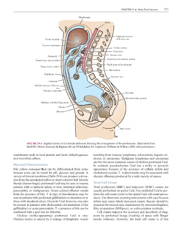
                                     FIGURE 29.6  Sagittal section o  the  emale abdomen showing the arrangement o  the peritoneum. (Reprinted  rom

                                     Snell RS. Clinical Anatomy By Regions, 8th ed, Philadelphia, PA: Lippincott Williams & Wilkins, 2008, with permission.)


                   constituents such as total protein and lactic dehydrogenase,                                                resulting  rom trauma, lymphoma, tuberculosis, hepatic cir-


                   and microbial culture.                                                                                      rhosis, or carcinoma. Malignant lymphoma and carcinoma

                                                                                                                               are the two most common causes o  chylous peritoneal f uid.
                   Physical Characteristics                                                                                    In  contrast,  pseudochylous  f uid  has  a  milky  or  greenish

                   Pale yellow abdominal   uid can be di  erentiated  rom urine                                                appearance because o  the presence o  cellular debris and


                   because urine can be tested  or pH, glucose, and protein. A                                                 cholesterol crystals. T is abnormality may be associated with
                   variety o  clinical conditions (  able 29.8) can produce a devia-                                           chronic e  usions produced by a wide variety o  causes.


                   tion  rom the anticipated yellow or straw-colored f uid. Grossly
                   bloody (hemorrhagic) peritoneal f uid may be seen in trauma                                                 Total Cell Count


                   patients with a ruptured spleen or liver, intestinal in arction,                                              otal erythrocyte (RBC) and leukocyte (WBC) counts are
                   pancreatitis, or malignancies. Green-colored e  usion results                                               usually per ormed on ascitic f uid. Use undiluted f uid to per-


                    rom the presence o  bile. T is type o  discoloration may be                                                 orm the cell count (re er to the spinal f uid cell count proce-
                   seen in patients with per orated  gallbladders or intestines or in                                          dure). Use electronic counting instruments with care because


                   those with duodenal ulcers. Greenish f uid, however, may also                                               debris may cause  alsely increased counts. Smears should be
                   be present in patients with cholecystitis (inf ammation o  the                                              prepared  or microscopic examination by cytocentri ugation,


                   gallbladder) or acute pancreatitis. T e presence o  bile can be                                              lter preparation (Millipore), or sedimentation methods.
                   con  rmed with a spot test  or bilirubin.                                                                        Cell counts improve the accuracy and speci  city o  diag-


                        Chylous  (milky-appearing)  peritoneal  f uid  is  rare.                                               nosis  by  peritoneal  lavage  (f ushing  o   space  with  Ringer
                   Chylous ascites is caused by a leakage o  lymphatic vessels                                                 lactate  solution).  However,  the  total  cell  count  is  o   less
   608   609   610   611   612   613   614   615   616   617   618