Page 611 - Clinical Hematology_ Theory _ Procedures ( PDFDrive )
P. 611

CHAPTER 29  ■  Body Fluid Analysis                        595


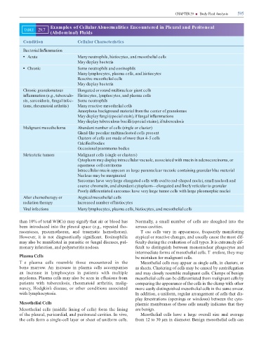
                                                Examples of Cellular Abnormalities Encountered in Pleural and Peritoneal
                       TABLE        29.7
                                                (Abdominal) Fluids


                       Condition                                         Cellular Characteristics


                       Bacterial In  ammation


                          Acute                                          Many neutrophils, histiocytes, and mesothelial cells
                                                                         May display bacteria


                          Chronic                                        Some neutrophils and eosinophils

                                                                         Many lymphocytes, plasma cells, and histiocytes

                                                                         Reactive mesothelial cells

                                                                         May display bacteria


                       Chronic granulomatous                             Elongated or round multinuclear giant cells

                       in  ammation (e.g., tuberculo- Histiocytes, lymphocytes, and plasma cells

                       sis, sarcoidosis, fungal infec- Some neutrophils

                       tions, rheumatoid arthritis)                      Many reactive mesothelial cells

                                                                         Amorphous background material from the center of granulomas

                                                                         May display fungi (special stain), if fungal in  ammations

                                                                         May display tuberculous bacilli (special stains), if tuberculosis


                       Malignant mesothelioma                            Abundant number of cells (single or cluster)

                                                                         Gland-like peculiar multinucleated cells present

                                                                         Clusters of cells are made of more than 4–5 cells

                                                                         Calci  ed bodies

                                                                         Occasional psammoma bodies

                       Metastatic tumors                                 Malignant cells (single or clusters)


                                                                         Cytoplasm may display intracellular vacuole, associated with mucin in adenocarcinoma, or
                                                                         squamous cell carcinoma


                                                                         Intracellular mucin appears as large paranuclear vacuole containing granular blue material
                                                                         Nucleus may be marginated


                                                                         Sarcomas have very large elongated cells with oval to rod-shaped nuclei, small nucleoli and
                                                                         coarse chromatin, and abundant cytoplasm—elongated and   nely reticular to granular


                                                                         Poorly differentiated sarcomas have very large tumor cells with large pleomorphic nuclei

                       After chemotherapy or                             Atypical mesothelial cells

                       radiation therapy                                 Increased number of histiocytes


                       Viral infections                                  Many lymphocytes, plasma cells, histiocytes, and mesothelial cells


                   than 10% o  total WBCs) may signi y that air or blood has                                                   Normally,  a small  number o  cells  are sloughed  into  the

                   been introduced into the pleural space (e.g., repeated tho-                                                 serous cavities.

                   racenteses,  pneumothorax,  and  traumatic  hemothorax).                                                         T ese  cells  vary  in  appearance,   requently  mani esting

                   However,  it  is  not  diagnostically  signi  cant.  Eosinophilia                                           atypical or reactive changes, and usually cause the most di -

                   may also be mani ested in parasitic or  ungal diseases, pul-                                                  culty during the evaluation o  cell types. It is extremely di -

                   monary in arction, and polyarteritis nodosa.                                                                  cult to distinguish between mononuclear phagocytes and

                                                                                                                               intermediate  orms o  mesothelial cells. T ere ore, they may
                   Plasma Cells                                                                                                be mistaken  or malignant cells.


                   Te  plasma  cells  resemble  those  encountered  in  the                                                         Mesothelial cells may appear as single cells, in clusters, or

                   bone  marrow.  An  increase  in  plasma  cells  accompanies                                                 as sheets. Clustering o  cells may be caused by centri ugation

                   an  increase  in  lymphocytes  in  patients  with  multiple                                                 and may closely resemble malignant cells. Clumps o  benign

                   myeloma. Plasma cells may also be seen in e  usions  rom                                                    mesothelial cells can be di  erentiated  rom malignant cells by

                   patients  with   tuberculosis,  rheumatoid  arthritis,  malig-                                              comparing the appearance o  the cells in the clump with other

                   nancy,  Hodgkin’s  disease,  or  other  conditions  associated                                              more easily distinguished mesothelial cells in the same smear.

                   with lymphocytosis.                                                                                         In addition, a uni orm, regular arrangement o  cells that dis-

                                                                                                                               play  enestrations (openings or windows) between the cyto-
                   Mesothelial Cells                                                                                           plasmic membranes o  these cells usually indicates that they


                   Mesothelial  cells (middle lining  o   cells)  orm the  lining                                              are benign.

                   o  the pleural, pericardial, and peritoneal cavities. In vivo,                                                   Mesothelial  cells  have  a  large  overall  size  and  average

                   the cells  orm a single-cell layer or sheet o  uni orm cells.                                                rom 12 to 30 µm in diameter. Benign mesothelial cells can
   606   607   608   609   610   611   612   613   614   615   616