Page 609 - Clinical Hematology_ Theory _ Procedures ( PDFDrive )
P. 609

CHAPTER 29  ■  Body Fluid Analysis                        593


                                                                                                                                    BOX  29.1


                                                                                                                                 Clinical Correlations (Pleural Fluid)


                                                                                                                                 TRANSUDATES

                                                                                                                                 Congestive heart  ailure

                                                                                                                                 Cirrhosis with ascites


                                                                                                                                 EXUDATES

                                                                                                                                 In ectious diseases

                                                                                                                                 Empyema

                                                                                                                                  uberculosis

                                                                                                                                 Malignant neoplasms

                                                                                                                                 Lymphoma

                                                                                                                                 Mesothelioma

                                                                                                                                 Pancreatitis

                                                                                                                                 Rheumatoid arthritis


                                                                                                                               Laboratory Analysis


                                                                                                                               Physical Characteristics

                                                                                                                                ransudates  are  usually  clear,  are  pale  yellow,  and  do  not

                                                                                                                               clot. In comparison, exudates can display a range o  colors

                                                                                                                               depending  on  the  associated  disorder  (  able  29.6).  Only

                                                                                                                               2 mL o  circulating blood in 1 L o  pleural f uid will pro-

                                                                                                                               duce a blood-tinged appearance. Very viscous f uids, clear

                                                                                                                               or bloody, are characteristic o  mesothelioma. In addition,

                                                                                                                               exudates may be cloudy or purulent and  requently clot on

                                                                                                                               standing because o  the presence o    brinogen.

                                                                                                                                    Specimen turbidity may be caused by lipids or result  rom
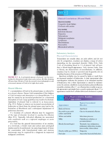
                   FIGURE 29.5  A. A substantial amount o  pleural f uid has accu-

                   mulated in this patient’s right chest cavity (arrow). B. A  er draining                                     an increased number o  leukocytes. A clear supernatant a  er
                   o   more than 100 mL o  f uid, the patient’s chest radiograph reveals                                       centri ugation indicates the presence o  an abundant number

                   a decreased amount o  f uid in the right chest cavity.                                                      o  leukocytes, but a white supernatant is caused by chylomi-
                                                                                                                               crons. In contrast, chyli orm or pseudochylous pleural e  usions

                                                                                                                               resemble a chylous e  ect. T ese e  usions have a milky or green-
                   Pleural Effusion
                                                                                                                               ish appearance and might have a pearly opalescent sheen. T is

                   T e accumulation o  f uid in the pleural space is re erred to                                               appearance results  rom cellular debris and cholesterol crystals.

                   as a pleural effusion. Excess f uid accumulates i  the balance

                   o  f uid  ormation and absorption is in disequilibrium. T is

                   may be caused by an increased production or a decreased                                                         TABLE        29.6        Representative Exudate

                   absorption o  f uid. Large quantities may need to be drained.                                                                            Appearance

                   Aspiration  o   pleural  f uid  is  re erred  to  as  thoracentesis                                                                                           Typical Associated

                   (Fig. 29.5). Failure to remove an increased accumulation o                                                      Appearance                                    Disorder

                   leukocytes or blood  rom the pleural space may lead to the

                    ormation o    brothorax and a subsequent impairment o                                                          Dark red-brown                                Amebiasis

                   pulmonary  unction.                                                                                             Greenish to greenish                          Classic rheumatoid effusion

                        Te  location  o   a  pleural  e  usion  may  be  suggestive                                                yellow and turbid

                   o   the  type  o   disorder  involved  in  causing  the  e  usion

                   (Box  29.1).    ypically,  le  -sided  e  usions  are  associated                                               Yellow and turbid                             Infectious process

                   with  conditions  such  as  a  ruptured  esophagus  or  acute                                                   Milky                                         Chylothorax (chylous or

                   pancreatitis.                                                                                                                                                 pseudochylous)

                        I  a  f uid has  the general  characteristics  o   an exudate,                                             Bloody (hemorrhagic)                          Traumatic tap, malignancy,

                   at a minimum, a Gram’s stain and culture and cytological                                                                                                      pulmonary infarction, trauma,

                     studies need to be per ormed. An open lung biopsy o   tissue                                                                                                pancreatitis, tuberculosis

                    or  examination  with  histochemical  stains  and  electron                                                    Clearly visible pus (WBCs)                    Empyema

                   microscopy  may  be  required   or  a  diagnosis  in  suspected

                   malignant conditions.                                                                                           Foul odor                                     Anaerobic bacterial infection
   604   605   606   607   608   609   610   611   612   613   614