
Page 604 - Clinical Hematology_ Theory _ Procedures ( PDFDrive )
P. 604
588 PART 8 ■ Fundamentals of Hematological Analysis
SPINAL FLUID: TOTAL LEUKOCYTE COUNT PROCEDURE (continued)
6. Soak the hemacytometer in a 10% bleach solution to disin- air bubbles. A er use, dispose o the hemacytometer
ect. Discard the capillary pipette and contaminated sup- in a biohazard container. I a di erent type o dispos-
plies in a biohazard bag. able hemacytometer is used, ollow the manu acturer’s
instructions and use the ormula provided to calculate
CALCULATIONS the cell count.
2. Clear specimens may be counted undiluted, provided no
10 overlapping o cells is seen on microscopic examination.
µ
9 × = 11 leukocytes L
/
9 When dilutions are required, calibrated automatic pipettes
are used. Dilutions are made with normal saline, mixed
(T ese calculations may need to be adjusted i the quantity by inversion, and loaded into the hemacytometer with a
o the specimen varies.)
micropipette. T e appropriate dilution actor must be used
in the calculation.
REFERENCE RANGES
3. Crystal violet stain can be used to acilitate the di erentia-
Normal CSF is crystal clear and colorless. No clots or RBCs tion o WBCs rom RBCs. Rinse a microhematocrit tube
should be observed. In addition, normal CSF has the vis- with crystal violet stain to coat the inside. Draw the f uid
cosity o water. into the coated microhematocrit tube, mix, and charge the
Normal values: 0 to 5 cells/µL or 0 to 5 × 10 /L (lymphocytes counting chamber.
6
and monocytes).
Some use a re erence value o 0 to 10/µL or 0 to 10 × 10 /L. CELLULAR ENUMERATION PROCEDURE NOTES
6
Neonates have a higher normal range, 0 to 30 mononuclear Sources of Error
cells × 10 /L. I the specimen is not examined promptly a er collection,
6
Values in children are comparable to those in adults. WBC lysis will give a alse impression o the number o
WBCs present. I a delay is anticipated, the specimen should
NOTES: be re rigerated.
1. A disposable, plastic hemacytometer may be used. T e Clotted specimens result in a alsely low cell count because
C-Chip DHC-N01 has a grid pattern and depth that are RBCs and WBCs will be trapped in the clot. In unusual cir-
the same as the Neubauer’s hemacytometer. T is all-in- cumstances, manual peripheral blood WBC or platelet counts
one unit does not require a coverslip. Use a micropipette may be needed. Unopettes or this procedure have been dis-
to load 10 µL o sample into the sample injection areas continued but Bioanalytic Gmg-H and Biomedical Polymers,
on either end o the chamber. T e chamber will ll by Gardner, MA, www.biomedicalpolymers.com, manu acture
capillary action. Be care ul to prevent introduction o substitutes.
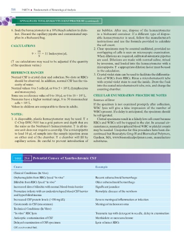
TABLE 29.4 Potential Causes of Xanthochromic CSF
Cause Example
Clinical Conditions (In Vivo)
Oxyhemoglobin from RBCs lysed “in vivo” Recent subarachnoid hemorrhage
Bilirubin from RBCs lysed “in vivo” Older subarachnoid hemorrhage
Increased direct bilirubin with normal blood-brain barrier Signi cant jaundice
Premature infants with an underdeveloped blood-CSF barrier Hemolytic disease of the newborn
and hyperbilirubinemia
Increased CSF protein levels (>150 mg/dL) Severe meningeal in ammation or infection
Carotenoids in CSF (uncommon) Meningeal melanosarcoma
Technical Conditions (In Vitro)
“In vitro” RBC lysis Traumatic tap with detergent in needle, delay in examination
Antiseptic contamination of CSF Merthiolate or mercurochrome
Delayed examination of CSF specimen Lysis of intact RBCs
CSF, cerebrospinal uid.