
Page 603 - Clinical Hematology_ Theory _ Procedures ( PDFDrive )
P. 603
CHAPTER 29 ■ Body Fluid Analysis 587
( able 29.4) and a delay in the examination o a speci- protein or gel ormation on standing due to an increased
men (which can cause a alse-positive result) can produce brinogen content.
xanthochromia.
Microscopic Exam ination: Cellular Enum eration
Viscosity Electronic cell counters are usually used to count cells in CSF.
Normal CSF has the viscosity o water. Clotting in CSF can Occasionally, total leukocyte cell counts on body f uids are
be caused by a variety o conditions, including increased per ormed manually.
SPINAL FLUID: TOTAL LEUKOCYTE COUNT PROCEDURE
PRINCIPLE
o enumerate the number o WBCs to assist in the devel-
opment o a di erential diagnosis (e.g., bacterial meningitis,
viral meningitis, ruptured brain abscess). W W
REAGENTS, SUPPLIES, AND EQUIPMENT
1. 10% acetic acid: Prepare by lling a 100-mL volumetric R R
f ask about hal ull with distilled water. Using a sa ety
bulb, pipette 10 mL o glacial acetic acid into the f ask.
Add distilled water to the calibration mark and mix. R
2. Wright-Giemsa or Wright’s stain or 1% methylene blue in
methyl alcohol: Prepare by weighing 1 g o methylene blue R R
and trans erring it to a 100-mL volumetric f ask. Dilute to
the calibration mark with methyl alcohol. Mix.
3. Small (12- × 75-mm) test tubes, Pasteur pipettes, rubber
bulb, and microscope slides W W
4. Neubauer’s hemacytometer
5. Centri uge, microscope, and immersion oil
6. Disin ectant solution
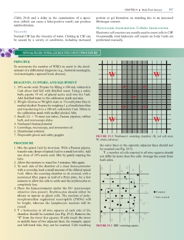
7. Disposable gloves and sa ety goggles FIGURE 29.2 Neubauer’s counting chamber. (R, red cell area;
W, white cell area.)
PROCEDURE
the outer lines or the opposite adjacent lines should not
1. Mix the spinal f uid by inversion. With a Pasteur pipette, be counted (see Fig. 29.3).
trans er nine drops o spinal f uid to a small test tube. Add T e number o cells counted in all nine squares should
one drop o 10% acetic acid. Mix by gently tapping the not di er by more than ve cells. Average the count rom
tube. both sides.
2. Allow this mixture to stand or 5 minutes. Mix again.
3. o each side o the chamber o a clean hemacytometer
with a coverslip, load a small amount o the diluted spinal
f uid. Allow the counting chamber to sit covered, with a
moistened lter paper in hal o a Petri plate, or a ew
minutes to allow the cells to settle and the erythrocytes to
completely lyse.
4. Place the hemacytometer under the 10× microscopic
objective (low power). Erythrocytes should either be Counted
absent or appear as ghost cells. he nucleus o poly- Not counted
morphonuclear segmented neutrophils (PMNs) will
be bright, whereas the lymphocyte nucleus will be
round.
5. T e leukocytes in all nine squares o each side o the
chamber should be counted (see Fig. 29.2). Remove the
“R” rom the inner ve squares. I cells touch the inner
or middle lines o two adjacent lines, or example, upper
and le -hand side, they can be counted. Cells touching FIGURE 29.3 RBC counting square.