
Page 614 - Clinical Hematology_ Theory _ Procedures ( PDFDrive )
P. 614
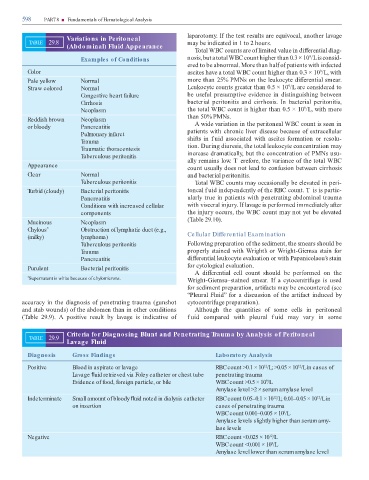
598 PART 8 ■ Fundamentals of Hematological Analysis
laparotomy. I the test results are equivocal, another lavage
Variations in Peritoneal
TABLE 29.8 may be indicated in 1 to 2 hours.
(Abdominal) Fluid Appearance
otal WBC counts are o limited value in di erential diag-
9
Examples f Conditions nosis, but a total WBC count higher than 0.3 × 10 /L is consid-
o
ered to be abnormal. More than hal o patients with in ected
Color ascites have a total WBC count higher than 0.3 × 10 /L, with
9
Pale yellow Normal more than 25% PMNs on the leukocyte di erential smear.
9
Straw colored Normal Leukocyte counts greater than 0.5 × 10 /L are considered to
Congestive heart failure be use ul presumptive evidence in distinguishing between
Cirrhosis bacterial peritonitis and cirrhosis. In bacterial peritonitis,
9
Neoplasm the total WBC count is higher than 0.5 × 10 /L, with more
than 50% PMNs.
Reddish brown Neoplasm
or bloody Pancreatitis A wide variation in the peritoneal WBC count is seen in
Pulmonary infarct patients with chronic liver disease because o extracellular
Trauma shi s in f uid associated with ascites ormation or resolu-
Traumatic thoracentesis tion. During diuresis, the total leukocyte concentration may
Tuberculous peritonitis increase dramatically, but the concentration o PMNs usu-
ally remains low. T ere ore, the variance o the total WBC
Appearance count usually does not lead to con usion between cirrhosis
Clear Normal and bacterial peritonitis.
Tuberculous peritonitis otal WBC counts may occasionally be elevated in peri-
Turbid (cloudy) Bacterial peritonitis toneal f uid independently o the RBC count. T is is partic-
Pancreatitis ularly true in patients with penetrating abdominal trauma
Conditions with increased cellular with visceral injury. I lavage is per ormed immediately a er
components the injury occurs, the WBC count may not yet be elevated
( able 29.10).
Mucinous Neoplasm
Chylous Obstruction of lymphatic duct (e.g.,
*
(milky) lymphoma) Cellular Differential Exam ination
Tuberculous peritonitis Following preparation o the sediment, the smears should be
Trauma properly stained with Wright’s or Wright-Giemsa stain or
Pancreatitis di erential leukocyte evaluation or with Papanicolaou’s stain
or cytological evaluation.
Purulent Bacterial peritonitis
A di erential cell count should be per ormed on the
* Supernatant is white because of chylomicrons. Wright-Giemsa–stained smear. I a cytocentri uge is used
or sediment preparation, arti acts may be encountered (see
“Pleural Fluid” or a discussion o the arti act induced by
accuracy in the diagnosis o penetrating trauma (gunshot cytocentri uge preparation).
and stab wounds) o the abdomen than in other conditions Although the quantities o some cells in peritoneal
( able 29.9). A positive result by lavage is indicative o fuid compared with pleural f uid may vary in some
Criteria for Diagnosing Blunt and Penetrating Trauma by Analysis of Peritoneal
TABLE 29.9
Lavage Fluid
Diagnosis Gross Findings Laboratory Analysis
Positive Blood in aspirate or lavage RBC count >0.1 × 10 /L; >0.05 × 10 /L in cases of
12
12
Lavage uid retrieved via Foley catheter or chest tube penetrating trauma
Evidence of food, foreign particle, or bile WBC count >0.5 × 10 /L
9
Amylase level >2 × serum amylase level
Indeterminate Small amount of bloody uid noted in dialysis catheter RBC count 0.05–0.1 × 10 /L; 0.01–0.05 × 10 /L in
12
12
on insertion cases of penetrating trauma
WBC count 0.001–0.005 × 10 /L
9
Amylase levels slightly higher than serum amy-
lase levels
Negative RBC count <0.025 × 10 /L
12
WBC count <0.001 × 10 /L
9
Amylase level lower than serum amylase level