
Page 635 - Clinical Hematology_ Theory _ Procedures ( PDFDrive )
P. 635
Digital microscopy Case studies
Describe the function of arti cial neural networks. Analyze and discuss the signi cance of the erythrocyte and leuko-
Explain the bene ts and advantages of digital microscopy. cyte histograms and the nomogram presented in the six case stud-
ies, answer the critical thinking group discussion questions, and
Instruments in coagulation studies conclude a diagnosis.
■ Describe the two most common types of instruments used in the
clinical laboratory for the detection of brin clots. NOTE:
■ Explain the principles of electromechanical and optical detection ■ indicates MLT and MLS core content
systems. indicates MLT (optional) and MLS advanced content
Describe the methodological principle of platelet aggregation.
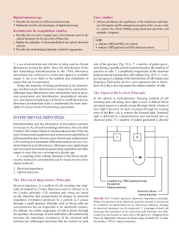
T e use o automation and robotics is being used in clinical side o the aperture (Fig. 30.1). T e number o pulses gener-
laboratories around the globe. Since the introduction o the ated during a speci c period is proportional to the number o
rst hematology instrumentation in the 1950s, laboratory particles or cells. T e amplitude (magnitude) o the electrical
automation has continued to evolve and expand in available pulse produced indicates the cell’s volume (Fig. 30.2). T e out-
assays. T ere is no limit to the number and complexity o put histogram is a display o the distribution o cell volume and
assays that can be automated. requency. Each pulse on the x-axis represents size in emto-
oday, the majority o testing per ormed in the hematol- liters ( L); the y-axis represents the relative number o cells.
ogy and hemostasis laboratories is impacted by automation.
Although larger laboratories have automation such as speci- The Optical Detection Principle
men preparation and identi cation, preanalytical automa-
tion, and digital morphology not ound in small laboratories, In the optical or hydrodynamic ocusing method o cell
laboratory pro essionals need to understand the basic prin- counting and cell sizing, laser light is used. A diluted blood
ciples o various levels o hematology automation. specimen passes in a steady stream through which a beam o
laser light is ocused. As each cell passes through the sensing
zone o the ow cell, it scatters the ocused light. Scattered
INSTRUMENTAL PRINCIPLES light is detected by a photodetector and converted into an
electrical pulse. T e number o pulses generated is directly
Instrumentation and the automation o procedures continue
to increase in the clinical hematology laboratory. Since the rst
Coulter Cell Counter Model A was introduced in the 1950s, the
types o automated equipment and instrumental capabilities o
instrumentation have become more diverse and sophisticated.
Cell counting and automated di erential analysis are now rou-
tinely ound in most laboratories. Microprocessor applications
have increased instrument programming capabilities and data
output in ways that were unimagined a decade ago.
T e counting o the cellular elements o the blood (eryth-
rocytes, leukocytes, and platelets) can be based on one o two
classic methods:
1. Electrical impedance
2. Optical detection
The Electrical Impedance Principle
Electrical impedance is a method o cell counting was origi-
nally developed by Coulter Electronics and is re erred to as
the Coulter principle. Cell counting and sizing are based
on the detection and measurement o changes in electrical
impedance (resistance) produced by a particle as it passes FIGURE 30.1 Coulter aperture: electronic impedance principle.
When the aperture o an electronic particle counter is immersed
through a small aperture. Particles such as blood cells are in a dilution o whole blood in an electrolyte solution, changes
nonconductive but are suspended in an electrically conduc- in electrical resistance can be measured. T e passage o each cell
tive diluent. As a dilute suspension o cells is drawn through increases the resistance o the electrical path between two elec-
the aperture, the passage o each individual cell momentarily trodes that are located on each side o the aperture. (Adapted rom
increases the impedance (resistance) o the electrical path Pierre R. Signi cant Advances in Hematology, Hialeah, FL: Coulter
between two submerged electrodes that are located on each Electronics, 1985:6, with permission.)