
Page 638 - Clinical Hematology_ Theory _ Procedures ( PDFDrive )
P. 638
622 PART 8 ■ Fundamentals of Hematological Analysis
react speci cally with the cellular component being stud- T e stained cells next pass through the laser beam. T e
ies such as reticulocytes, peroxidase enzyme, or DNA con- laser activates the dye and the cell f uoresces. T e interaction
tent. Fluorescent dyes include acridine orange, thiof avin , between each cell and the laser beam provides the ollowing
pyronin Y, f uorescein isothiocyanate (FI C), and phycoery- two types o in ormation:
thrin (PE). FI C and PE are used when dual color analysis 1. T e amount o light scattered by each cell hit by the laser
is desired. beam
Many f ow cytometric assays use direct immunof uores-
cence staining with f uorochrome-conjugated monoclonal 2. T e intensity o the f uorescence emitted by labeled anti-
bodies bound to antigens on the di erent types o sus-
antibodies to identi y cellular characteristics. Fluorochromes pended cells
are molecules that absorb light o one wavelength and emit
light o a higher wavelength. Fluorochromes are covalently Although the f uorescence is emitted throughout a
bonded to monoclonal antibody molecules. T is provides 360-degree circle, it is usually collected via optical sensors
a mechanism that allows or the determination by the f ow located at 90 degrees relative to the laser beam. T e f uores-
cytometer i a labeled antibody has bound to the cell sur ace. cence in ormation is then transmitted to a computer. Flow
Each f uorochrome has a maximal excitation wavelength cytometry per orms f uorescence analysis on single cells at
at or near the wavelength o the laser and has a characteris- rates up to 50,000 cells/minute. T e computer is the heart o
tic emission spectrum. T e f uorochrome, excited by the laser the instrument; it controls all decisions regarding data col-
light, will f uoresce at a longer wavelength. Fluorescein emits lection, analysis, and cell sorting.
a green f uorescence, PE emits orange, and peridinin chloro-
phyll protein or PE coupled to cyanin 5 emits a red f uores- The Basis of Cellular Identi cation
cence. An argon laser, which produces blue light, is the most
commonly used laser. Some instruments add a red helium- One o the major advantages o f ow cytometry is that more
neon laser, and occasionally, a mercury arc lamp is substituted. than one measurement can be made on every cell during
Some f ow cytometers have a second laser that can excite the ew milliseconds that the cell spends passing through
other f uorochromes. Like the side-scattered blue light, all the laser beam. Each cell can be optically measured or the
o these f uorescent signals pass through the objective set at intensity o scattered light.
90 degrees to the incident laser light. T e number o colors Te cellular light scatter patterns can be used to iden-
in f ow cytometry output re ers to the number o individual ti y cells. Both intrinsic and extrinsic properties o cells
f uorochrome-labeled antibodies used simultaneously in a can be analyzed by f ow cytometry. Intrinsic properties
given reaction tube. For example, mixing a cell suspension include orward- and right-angle light scatter, which cor-
with a combination o antibodies labeled with two f uoro- relate with size and granularity o a cell, respectively. T is
chromes is re erred to two-color f ow cytometry. data output does not require addition o dyes or stains or
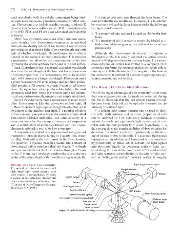
A suspension o stained cells is pressurized using gas and detection. In contrast, extrinsic properties rely on the bind-
transported through plastic tubing to a quarts f ow cham- ing o various probes to the cells. T e scattered light passes
ber (Fig. 30.4) within the instrument. In the f ow chamber, through a variety o lters and lenses and is then measured
the specimen is injected through a needle into a stream o by photomultiplier tubes, which convert the light signals
physiological saline solution called the sheath. T e sheath into electronic signals or computer analysis. Light scat-
and specimen both exit the f ow chamber through a 75-µm tered along the axis o the laser beam is “ orward scatter,”
ori ce. T is laminar f ow design con nes the cells to the very and light scattered perpendicular to the axis is “side scat-
center o the saline sheath with the cells moving in single le. ter” or “orthogonal scatter.” Forward scatter is roughly
FIGURE 30.4 Laser f ow cytometry.
Te optical detection o orward- and
right-angle light scatter using a laser
light source is accomplished by using
a sensor as the cells pass through the
beam under conditions o laminar f ow.
(Courtesy o Ortho Diagnostic Systems,
Westwood, MA, 1985.)