
Page 637 - Clinical Hematology_ Theory _ Procedures ( PDFDrive )
P. 637
CHAPTER 30 ■ Instrumentation in Hematology 621
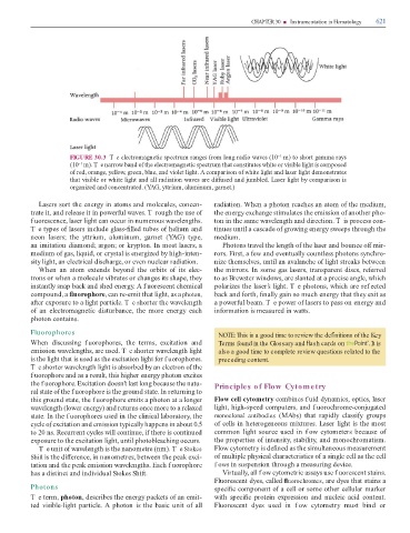
FIGURE 30.3 T e electromagnetic spectrum ranges rom long radio waves (10 m) to short gamma rays
−1
(10 m). T e narrow band o the electromagnetic spectrum that constitutes white or visible light is composed
−1
o red, orange, yellow, green, blue, and violet light. A comparison o white light and laser light demonstrates
that visible or white light and all radiation waves are di used and jumbled. Laser light by comparison is
organized and concentrated. (YAG, yttrium, aluminum, garnet.)
Lasers sort the energy in atoms and molecules, concen- radiation. When a photon reaches an atom o the medium,
trate it, and release it in power ul waves. T rough the use o the energy exchange stimulates the emission o another pho-
f uorescence, laser light can occur in numerous wavelengths. ton in the same wavelength and direction. T is process con-
T e types o lasers include glass- lled tubes o helium and tinues until a cascade o growing energy sweeps through the
neon lasers; the yttrium, aluminum, garnet (YAG) type, medium.
an imitation diamond; argon; or krypton. In most lasers, a Photons travel the length o the laser and bounce o mir-
medium o gas, liquid, or crystal is energized by high-inten- rors. First, a ew and eventually countless photons synchro-
sity light, an electrical discharge, or even nuclear radiation. nize themselves, until an avalanche o light streaks between
When an atom extends beyond the orbits o its elec- the mirrors. In some gas lasers, transparent discs, re erred
trons or when a molecule vibrates or changes its shape, they to as Brewster windows, are slanted at a precise angle, which
instantly snap back and shed energy. A f uorescent chemical polarizes the laser’s light. T e photons, which are ref ected
compound, a uorophore, can re-emit that light, as a photon, back and orth, nally gain so much energy that they exit as
a er exposure to a light particle. T e shorter the wavelength a power ul beam. T e power o lasers to pass on energy and
o an electromagnetic disturbance, the more energy each in ormation is measured in watts.
photon contains.
Fluorophores NOTE: This is a good time to review the de nitions of the Key
When discussing f uorophores, the terms, excitation and Terms found in the Glossary and ash cards on . It is
emission wavelengths, are used. T e shorter wavelength light also a good time to complete review questions related to the
is the light that is used as the excitation light or f uorophores. preceding content.
T e shorter wavelength light is absorbed by an electron o the
fuorophore and as a result, this higher energy photon excites
the f uorophore. Excitation doesn’t last long because the natu- Principles of Flow Cytometry
ral state o the f uorophore is the ground state. In returning to
this ground state, the f uorophore emits a photon at a longer Flow cell cytometry combines f uid dynamics, optics, laser
wavelength (lower energy) and returns once more to a relaxed light, high-speed computers, and f uorochrome-conjugated
state. In the f uorophores used in the clinical laboratory, the monoclonal antibodies (MAbs) that rapidly classi y groups
cycle o excitation and emission typically happens in about 0.5 o cells in heterogeneous mixtures. Laser light is the most
to 20 ns. Recurrent cycles will continue, i there is continued common light source used in f ow cytometers because o
exposure to the excitation light, until photobleaching occurs. the properties o intensity, stability, and monochromatism.
Te unit o wavelength is the nanometre (nm). Te Stokes Flow cytometry is de ned as the simultaneous measurement
Shift is the di erence, in nanometres, between the peak exci- o multiple physical characteristics o a single cell as the cell
tation and the peak emission wavelengths. Each f uorophore f ows in suspension through a measuring device.
has a distinct and individual Stokes Shi . Virtually, all f ow cytometric assays use f uorescent stains.
Fluorescent dyes, called uorochromes, are dyes that stains a
Photons speci c component o a cell or some other cellular marker
Te term, photon, describes the energy packets o an emit- with speci c protein expression and nucleic acid content.
ted visible-light particle. A photon is the basic unit o all Fluorescent dyes used in f ow cytometry must bind or