Page 688 - Clinical Hematology_ Theory _ Procedures ( PDFDrive )
P. 688

672            PART 8  ■  Fundamentals of Hematological Analysis


                       HEMATOLOGY PROCEDURES (continued)


               Clinical Applications


               In most cases, leukocyte counts are only per orme   manu-

               ally when there are extremely low total leukocyte counts.

               T e total leukocyte count in whole bloo   specimens can be

                 ecrease   or increase     ue to a variety o    isor  ers.


               Selecte   quantitative leukocyte   isor  ers

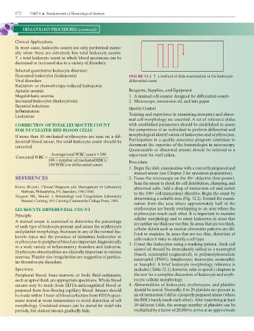
               Decrease   leukocytes (leukopenia)                                                                           FIGURE 32.2  T e metho   o  sli  e examination in the leukocyte

               Viral   isor  ers                                                                                             i  erential count.

               Ra  iation- or chemotherapy-in  uce   leukopenia

               Aplastic anemia                                                                                             Reagents, Supplies, and Equipment

               Megaloblastic anemia                                                                                        1.  A manual cell counter   esigne    or   i  erential counts

               Increase   leukocytes (leukocytosis)                                                                        2.  Microscope, immersion oil, an   lens paper

               Bacterial in ections                                                                                        Quality Control

               In  ammation

               Leukemias                                                                                                    raining an   experience in examining immature an   abnor-
                                                                                                                           mal cell morphology are essential. A set o  re erence sli  es

               CORRECTION OF TOTAL LEUKOCYTE COUNT                                                                         with establishe   parameters shoul   be establishe   to assess


               FOR NUCLEATED RED BLOOD CELLS                                                                               the competence o  an in  ivi  ual to per orm   i  erential an

               I  more than 10 nucleate   erythrocytes are seen on a   i -                                                 morphological i  entif cation o  leukocytes an   erythrocytes.

                erential bloo   smear, the total leukocyte count shoul   be                                                Participation in a quality assurance program continues to

               correcte  .                                                                                                   ocument the expertise o  the hematologist in microscopy.
                                                                                                                           Questionable or abnormal smears shoul   be re erre   to a

                                                 Averagetotal WBC count                      ×100                          supervisor  or verif cation.
                Correcte  WBC =
                                               100    +  number o  nucleate  RRBCs/                                        Procedure

                                               100    WBCsin   i  erential count
                                                                                                                           1.  Begin the sli  e examination with a correctly prepare   an

                                                                                                                                staine   smear (see Chapter 2  or specimen preparation).

               REFERENCES                                                                                                  2.  Focus the microscope on the 10× objective (low power).

                                                                                                                                Scan the smear to check  or cell   istribution, clumping, an
               Henry JB (e  .). Clinical Diagnosis and Management by Laboratory                                                 abnormal cells. A     a   rop o  immersion oil an   switch

                     Methods, Phila  elphia, PA: Saun  ers, 1984:1444.                                                          to the 100× (oil immersion) objective. Begin the count by

                urgeon  ML,  Ben  er  J.  Hematology  and  Coagulation  Laboratory                                               etermining a suitable area (Fig. 32.2). Exten  the exami-

                     Manual, Corning, NY: Corning Community College Press, 1985.
                                                                                                                                nation   rom  the  area  where  approximately  hal   o   the

               LEUKOCYTE DIFFERENTIAL COUNT                                                                                     erythrocytes are barely overlapping to an area where the


               Principle                                                                                                        erythrocytes touch each other. It is important to examine

               A staine   smear is examine   to   etermine the percentage                                                       cellular morphology and to count leukocytes in areas that
                                                                                                                                are neither too thick nor too thin. In areas that are too thick,
               o  each type o  leukocyte present an   assess the erythrocyte                                                    cellular   etails such as nuclear chromatin patterns are   i -

               an   platelet morphology. Increases in any o  the normal leu-                                                    fcult to examine. In areas that are too thin,   istortion o

               kocyte types an   the presence o  immature leukocytes or                                                         cells makes it risky to i  enti y a cell type.

               erythrocytes in peripheral bloo   are important   iagnostically                                             3.  Count the leukocytes using a tracking pattern. Each cell

               in a wi  e variety o  in  ammatory   isor  ers an   leukemia.                                                    i  entif e   shoul   be imme  iately tallie   as a neutrophil

               Erythrocyte abnormalities are clinically important in various                                                    (ban  ), neutrophil (segmente  ), or polymorphonuclear

               anemias. Platelet size irregularities are suggestive o  particu-                                                 neutrophil (PMN); lymphocyte; monocyte; eosinophil;

               lar thrombocyte   isor  ers.
                                                                                                                                or basophil. A brie  leukocyte morphology re erence is

               Specimen                                                                                                         inclu  e   (  able 32.1); however, re er to specif c chapters in

               Peripheral bloo  , bone marrow, or bo  y   ui   se  iments,                                                      the text  or a complete   iscussion o  leukocyte an   eryth-

               such as spinal   ui  , are appropriate specimens. Whole bloo                                                     rocyte cellular morphology.

               smears may be ma  e  rom ED  A-anticoagulate   bloo   or                                                    4.  Abnormalities o  leukocytes, erythrocytes, an   platelets

               prepare    rom  ree-  owing capillary bloo  . Smears shoul                                                       shoul   be note  . Normally, 8 to 20 platelets are present in

               be ma  e within 1 hour o  bloo   collection  rom ED  A speci-                                                    an oil immersion f el   in a properly prepare   smear (where

               mens store   at room temperature to avoi     istortion o  cell                                                   the RBCs barely touch each other). A  er  examining at least

               morphology. Unstaine   smears can be store    or in  ef nite                                                     10   i  erent f el  s, the average number o  platelets can be

               perio  s, but staine   smears gra  ually  a  e.                                                                  multiplie   by a  actor o  20,000 to arrive at an approximate
   683   684   685   686   687   688   689   690   691   692   693