Page 689 - Clinical Hematology_ Theory _ Procedures ( PDFDrive )
P. 689

CHAPTER 32  ■  Laboratory Manual: Manual Procedures in Hematology                                         673


                             HEMATOLOGY PROCEDURES (continued)


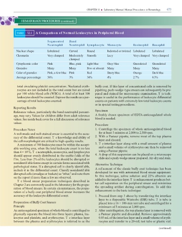
                       TABLE         32.1       A Comparison of Normal Leukocytes in Peripheral Blood


                                                                Segmented                    Band

                                                                Neutrophil                   Neutrophil Lymphocyte Monocyte                                                      Eosinophil                Basophil


                       Nuclear shape                            Lobulated                    Curved                 Round                      Indented or twisted Lobulated                               Lobulated

                       Chromatin                                Very clumped                 Moderately             Smooth                     Lacy                              Very clumped Very clumped


                                                                                             clumped

                       Cytoplasmic color                        Pink                         Blue, pink             Light blue                 Gray-blue                         Granulated                Granulated


                       Granules                                 Many                         Many                   Few or absent              Many                              Many                      Many


                       Color of granules                        Pink, a few blue Pink                               Red                        Dusty blue                        Orange                    Dark blue

                       Average percentage                       56%                          3%                     34%                        4%                                2.7%                      0.3%


                        total circulating platelet concentration. Nucleate   eryth-                                            bu  y coat. I  this layer o  concentrate   cells is remove   by

                        rocytes are not inclu  e   in the total count but are note                                             pipetting, push-we  ge–type smears can subsequently be pre-

                        per 100 white bloo   cells (WBCs). A total o  at least 100                                             pare   an   staine    or microscopic examination. T is tech-

                        leukocytes shoul   be counte  . Express the results as a per-                                          nique is use ul in the per ormance o  leukocyte   i  erential

                        centage o  total leukocytes counte  .                                                                  counts on patients with extremely low total leukocyte counts

                                                                                                                               or in special testing proce  ures.

                   Reporting Results

                   Re erence values, particularly the ban   neutrophil percent-                                                Specimen

                   age, may vary. Values  or chil  ren   i  er  rom a  ult re erence                                           A  reshly   rawn specimen o  ED  A-anticoagulate   whole

                   values. See insi  e back cover  or a  ull   iscussion o  re erence                                          bloo   is nee  e  .

                   values.
                                                                                                                               Procedure


                   Procedure Notes                                                                                             1.  Centri uge the specimen o  whole anticoagulate   bloo

                   A well-ma  e an   well-staine   smear is essential to the accu-                                                   or at least 5 minutes at 2,000 to 2,500 rpm.

                   racy o  the   i  erential count. T e knowle  ge an   ability o                                              2.  With a Pasteur pipette, remove most o  the top plasma

                   the cell morphologist are critical to high-quality results.                                                      layer an     iscar  .

                        A minimum o  300 leukocytes must be within the accept-                                                 3.  T e inter ace layer along with a small amount o  plasma

                   able working area, when the total leukocyte count is no less                                                     an   a small volume o  erythrocytes can then be remove

                   than 4 × 10 /L. T e neutrophils, monocytes, an   lymphocytes                                                     using a Pasteur pipette.
                                     9
                   shoul   appear evenly   istribute   in the usable f el  s o  the                                            4.  A   rop o  this suspension can be place   on a microscope

                   f lm. Less than 2% o  the leukocytes shoul   be   isrupte   or                                                   sli  e an   a push-we  ge smear prepare  . Air-  ry an   stain.

                   noni  entif able  orms except in certain  orms associate   with                                             Alternative Technique

                   pathological states. I  a   isrupte   cell is clearly i  entif able,

                   inclu  e it in the   i  erential count. Classi y noni  entif able                                           A ref nement o  the classic bu  y coat technique has been

                     isrupte   cells (smu  ges or baskets) as “other,” an   note them                                            evelope    or use with automate   bloo   smear equipment.

                   on the report i  more than a  ew are observe  .                                                             In  this  technique,  saline  solution  an    22%  albumin  are

                        Te bloo   smear preparation techniques    escribe   in                                                 a    e   to the inter ace layer. T is enhancement pro  uces bet-

                   Chapter 2 are commonly use   in the laboratory  or the prepa-                                               ter cell separation on the peripheral smear an   minimizes

                   ration o  bloo   smears. In certain circumstances, the prepa-                                               the sprea  ing arti act   uring centri ugation.   o a     this

                   ration o  a bu  y coat peripheral bloo   smear increases the                                                enhancement to the basic technique:

                   accuracy o  the leukocyte   i  erential count.                                                              1.  Procee    rom step 3 above by trans erring the inter ace

                                                                                                                                    layer to a   isposable Wintrobe (ESR) tube. T is tube is

                   Preparation of Buffy Coat Smears                                                                                 place   into a 16 × 100-mm test tube an   centri uge    or a

                   Principle                                                                                                        minimum o  5 minutes at 2,000 rpm.

                   An anticoagulate   specimen o  whole bloo   is centri uge   to                                              2.  A  er centri ugation, the top plasma layer is remove   with

                   physically separate the bloo   into three layers: plasma, leu-                                                   a Pasteur pipette an     iscar  e  . Remove approximately

                   kocytes an   platelets, an   erythrocytes. T e inter ace layer                                                   0.03 mL o  the inter ace layer an   a small volume o  eryth-

                   between the plasma an   erythrocytes is re erre   to as the                                                      rocytes an   trans er to a 20-mL test tube or plastic vial.


                                                                                                                                                                                                                    (continued)
   684   685   686   687   688   689   690   691   692   693   694