
Page 692 - Clinical Hematology_ Theory _ Procedures ( PDFDrive )
P. 692
676 PART 8 ■ Fundamentals of Hematological Analysis
HEMATOLOGY PROCEDURES (continued)
Calculation
=
No.o erythrocytes average
ra itional Formula:
total o RBCscounnte in 5squares
× ilution correction actor
× volumecorrection aactor
1. Five o the 25 squares in the large 1-mm square are counte .
2. T e specimen ilution actor is 200.
3. T e volume correction actor is 50. T is number rep-
resents the total volume o the f ve squares reporte in
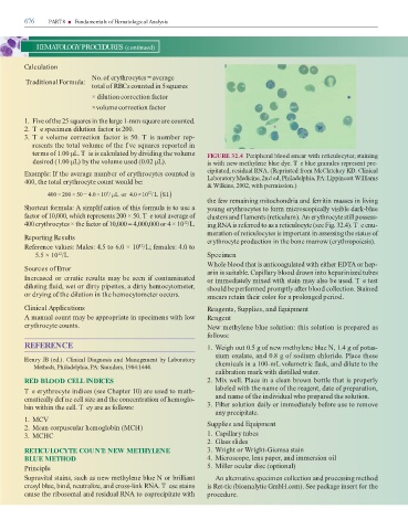
terms o 1.00 µL. T is is calculate by ivi ing the volume FIGURE 32.4 Peripheral bloo smear with reticulocytes; staining
esire (1.00 µL) by the volume use (0.02 µL). is with new methylene blue ye. T e blue granules represent pre-
Example: I the average number o erythrocytes counte is cipitate , resi ual RNA. (Reprinte rom McClatchey KD. Clinical
Laboratory Medicine, 2n e , Phila elphia, PA: Lippincott Williams
400, the total erythrocyte count woul be:
& Wilkins, 2002, with permission.)
6
12
×
400 200 50 4 0 10 / µLor 4 0 10 / L ( .SI. )
×
=
.
×
.
×
the ew remaining mitochon ria an erritin masses in living
Shortcut ormula: A simplif cation o this ormula is to use a young erythrocytes to orm microscopically visible ark-blue
actor o 10,000, which represents 200 × 50. T e total average o clusters an f laments (reticulum). An erythrocyte still possess-
400 erythrocytes × the actor o 10,000 = 4,000,000 or 4 × 10 /L. ing RNA is re erre to as a reticulocyte (see Fig. 32.4). T e enu-
12
meration o reticulocytes is important in assessing the status o
Reporting Results erythrocyte pro uction in the bone marrow (erythropoiesis).
Re erence values: Males: 4.5 to 6.0 × 10 /L; emales: 4.0 to
12
5.5 × 10 /L Specimen
12
Whole bloo that is anticoagulate with either ED A or hep-
Sources of Error arin is suitable. Capillary bloo rawn into heparinize tubes
Increase or erratic results may be seen i contaminate or imme iately mixe with stain may also be use . T e test
iluting ui , wet or irty pipettes, a irty hemocytometer, shoul be per orme promptly a er bloo collection. Staine
or rying o the ilution in the hemocytometer occurs. smears retain their color or a prolonge perio .
Clinical Applications Reagents, Supplies, and Equipment
A manual count may be appropriate in specimens with low Reagent
erythrocyte counts. New methylene blue solution: this solution is prepare as
ollows:
REFERENCE 1. Weigh out 0.5 g o new methylene blue N, 1.4 g o potas-
sium oxalate, an 0.8 g o so ium chlori e. Place these
Henry JB (e .). Clinical Diagnosis and Management by Laboratory
Methods, Phila elphia, PA: Saun ers, 1984:1444. chemicals in a 100-mL volumetric ask, an ilute to the
calibration mark with istille water.
RED BLOOD CELL INDICES 2. Mix well. Place in a clean brown bottle that is properly
Te erythrocyte in ices (see Chapter 10) are use to math- labele with the name o the reagent, ate o preparation,
ematically ef ne cell size an the concentration o hemoglo- an name o the in ivi ual who prepare the solution.
bin within the cell. T ey are as ollows: 3. Filter solution aily or imme iately be ore use to remove
any precipitate.
1. MCV
2. Mean corpuscular hemoglobin (MCH) Supplies and Equipment
3. MCHC 1. Capillary tubes
2. Glass sli es
RETICULOCYTE COUNT: NEW METHYLENE 3. Wright or Wright-Giemsa stain
BLUE METHOD 4. Microscope, lens paper, an immersion oil
Principle 5. Miller ocular isc (optional)
Supravital stains, such as new methylene blue N or brilliant An alternative specimen collection an processing metho
cresyl blue, bin , neutralize, an cross-link RNA. T ese stains is Ret-tic (bioanalytic GmbH.com). See package insert or the
cause the ribosomal an resi ual RNA to coprecipitate with proce ure.