
Page 696 - Clinical Hematology_ Theory _ Procedures ( PDFDrive )
P. 696
680 PART 8 ■ Fundamentals of Hematological Analysis
SPECIAL HEMATOLOGY PROCEDURES
ACIDIFIED SERUM LYSIS TEST: HAM METHOD
Principle
Erythrocytes are incubate with resh an heate serum to test
or hemolysis. Weak aci is use in specif c serum cell mix-
tures to maximize hemolytic activity. T e presence o hemo-
lysis, epen ing on the test con itions, may be observe in
cases o antibo y-sensitize coate erythrocytes, spherocytes,
or paroxysmal nocturnal hemoglobinuria (PNH).
T is proce ure, in CLSI ormat, is provi e on this book’s
companion Web site at http://thepoint.lww.com/ urgeon6e.
BONE MARROW EXAMINATION
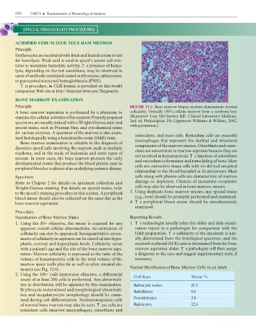
Principle FIGURE 32.6 Bone marrow biopsy sections emonstrate normal
A bone marrow aspiration is per orme by a physician to cellularity. Virtually 100% cellular marrow rom a newborn boy.
examine the cellular activities o the marrow. Properly prepare (Reprinte rom McClatchey KD. Clinical Laboratory Medicine,
specimens are usually staine with a Wright-Giemsa stain an 2n e , Phila elphia, PA: Lippincott Williams & Wilkins, 2002,
special stains, such as Prussian blue, an cytochemical stains with permission.)
or various enzymes. A specimen o the marrow is also exam- osteoclasts, an mast cells. Reticulum cells are peace ul
ine histologically using a hematoxylin-eosin (H&E) stain. macrophages that represent the skeletal an structural
Bone marrow examination is valuable in the iagnosis o
isor ers specif cally involving the marrow, such as multiple components o the marrow sinuses. Osteoblasts an osteo-
clasts are uncommon in marrow aspirates because they are
myeloma, an in the stu y o leukemias an some types o not involve in hematopoiesis. T e unction o osteoblasts
anemia. In most cases, the bone marrow presents the early an osteoclasts is ormation an remo eling o bone. Mast
evelopmental events that pro uce the bloo picture seen in cells are connective tissue cells with no ef ne ancestral
peripheral bloo or evi ence o an un erlying systemic isease.
relationship to the bloo basophil or its precursors. Mast
Specimen cells along with plasma cells are characteristic o marrow
Re er to Chapter 2 or etails on specimen collection an amage or epletion. Clusters o metastatic neoplastic
Wright-Giemsa staining. For etails on special stains, re er cells may also be observe in bone marrow smears.
to the specif c staining proce ure in this section. A peripheral 3. Using uplicate bone marrow smears, any special stains
bloo smear shoul also be collecte on the same ay as the (e.g., iron) shoul be promptly per orme an examine .
bone marrow aspiration. 4. T e peripheral bloo smear shoul be simultaneously
examine .
Procedure
Examination of Bone Marrow Slides Reporting Results
1. Using the 10× objective, the smear is scanne or any 1. T e technologist usually re ers the sli es an sli e exami-
apparent overall cellular abnormalities. An estimation o nation report to a pathologist or comparison with the
cellularity can also be appraise . Semiquantitative assess- H&E preparation. T e cellularity o the specimen is usu-
ments o cellularity in aspirates can be classif e into hypo- ally etermine rom the histological specimen, an the
plastic, normal, an hyperplastic levels. Cellularity varies myeloi -erythroi (M/E) ratio is etermine rom the bone
with a patient’s age an the site o the bone marrow aspi- marrow aspiration sli es. T e pathologist will then assign
ration. Marrow cellularity is expresse as the ratio o the a iagnosis to the case an suggest supplementary tests, i
volume o hematopoietic cells to the total volume o the necessary.
marrow space (cells plus at as well as other stromal ele-
ments) (see Fig. 32.6). Normal Distribution of Bone Marrow Cells in an Adult
2. Using the 100× (oil) immersion objective, a i erential Cell Type Mean %
count o at least 200 cells is per orme . Any abnormali-
ties in istribution will be apparent by this examination. Rubricytic series 21.5
Erythrocyte maturational an morphological abnormali- Rubriblasts 0.6
ties an megakaryocyte morphology shoul be exam-
ine uring cell i erentiation. Nonhematopoietic cells Prorubricytes 2.0
o normal bone marrow may also be seen. T ese cells are Rubricytes 12.4
reticulum cells (marrow macrophages), osteoblasts an