Page 697 - Clinical Hematology_ Theory _ Procedures ( PDFDrive )
P. 697

CHAPTER 32  ■  Laboratory Manual: Manual Procedures in Hematology                                         681


                             SPECIAL HEMATOLOGY PROCEDURES (continued)


                                                                                                                               nucleoti  es, when activate   with long-wave (340 to 370 nm)
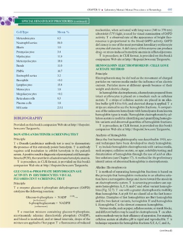
                       Cell Type                                             Mean %
                                                                                                                               ultraviolet (UV) light, is use    or visual examination o  G6PD


                       Metarubricytes                                        6.5                                               activity. T e observe   rate o  the appearance o  bright   uo-
                                                                                                                               rescence is proportional to the bloo   G6PD activity. G6PD
                       Neutrophil series                                     56.0
                                                                                                                                ef ciency is one o  the most prevalent here itary erythrocyte
                       Blasts                                                1.0                                               enzyme   ef ciencies. A   ef ciency o  this enzyme can pro  uce


                       Promyelocytes                                         3.4                                                rug- or stress-in  uce  hemolytic anemia in a  icte  persons.


                       Myelocytes                                            11.9                                                   T is proce  ure, in CLSI  ormat, is provi  e   on this book’s
                                                                                                                               companion Web site at http://thepoint.lww.com/  urgeon6e.
                       Metamyelocytes                                        18.0


                       Bands                                                 11.0                                              HEMOGLOBIN ELECTROPHORESIS: CELLULOSE

                                                                                                                               ACETATE METHOD
                       PMNs                                                  10.7
                                                                                                                               Principle
                       Eosinophil series                                     3.2
                                                                                                                               Electrophoresis may be   ef ne   as the movement o  charge
                       Basophils                                             <0.1                                              particles on various me  ia un  er the in  uence o  an electric


                       Lymphocytes                                           15.8                                              current. Particles move at   i  erent spee  s because o  their


                       Monocytes                                             1.8                                               weight an   electric charge.
                                                                                                                                    In hemoglobin electrophoresis, a hemolysate prepare    rom
                       Megakaryocytes                                        <0.1
                                                                                                                               intact erythrocytes is place   on a me  ium such as cellulose
                       Reticulum cells                                       0.3
                                                                                                                               acetate. T e strips o  cellulose acetate are place   in an alka-

                       Plasma cells                                          1.8                                               line bu  er (pH 8.0 to 8.6), an   electrical charge is applie  . T e


                       M/E ratio                                             2.5:1                                             strips are staine   to see the hemoglobin  ractions. A compari-
                                                                                                                               son o  the unknown hemolysate with hemolysates  rom known

                                                                                                                               hemoglobin types is ma  e. Hemoglobin electrophoresis by cel-

                   BIBLIOGRAPHY                                                                                                lulose acetate is use ul in i  enti ying an   quanti ying hemoglo-

                                                                                                                               bin variants an   abnormal quantities o  hemoglobin  ractions.

                   Provi  e   on this book’s companion Web site at http://thepoint.                                                 Tis proce  ure, in CLSI  ormat, is provi  e   on this book’s

                   lww.com/  urgeon6e.                                                                                         companion Web site at http://thepoint.lww.com/  urgeon6e.


                   DONATH-LANDSTEINER SCREENING TEST                                                                           Analysis of Hemoglobin

                   Principle                                                                                                   Since the f rst hemoglobinopathy was   escribe   in 1910, sev-


                   T e Donath-Lan  steiner antibo  y test is use   to   emonstrate                                             eral techniques have been   evelope   to stu  y hemoglobin.

                   the presence o  this extremely potent hemolysin. T is antibo  y                                             Tey inclu  e hemoglobin electrophoresis with various me  ia,

                   requires col   incubation to exhibit hemolysis in the patient’s                                             such as paper, cellulose acetate, or agar; solubility testing; an

                   serum. A positive result is   iagnostic o  paroxysmal col   hemoglo-                                          enaturation o  hemoglobin through the use o  aci   or alka-

                   binuria (PCH), the rarest  orm o  autoimmune hemolytic anemia.                                              line solutions (see Chapter 17). A metho    or the preliminary

                        T is proce  ure, in CLSI  ormat, is provi  e   on this book’s                                          i  entif cation o  abnormal hemoglobin is electrophoresis.

                   companion Web site at http://thepoint.lww.com/  urgeon6e.
                                                                                                                               Alkaline Electrophoresis

                   GLUCOSE-6-PHOSPHATE DEHYDROGENASE                                                                           T is metho   o  separating hemoglobin  ractions is base   on

                   ACTIVITY IN ERYTHROCYTES: VISUAL                                                                            the principle that hemoglobin molecules in an alkaline solu-

                   FLUORESCENT SCREENING TEST                                                                                  tion have a net negative charge an   move towar   the ano  e in


                   Principle                                                                                                   an electrophoretic system. T is screening proce  ure can sep-

                   Te enzyme glucose-6-phosphate   ehy  rogenase (G6PD)                                                        arate hemoglobins A, F, S, an   C an   other variant hemoglo-

                   catalyzes the  ollowing reaction:                                                                           bins (Fig. 32.7). T ose with a greater electrophoretic mobility
                                                                                                                               than hemoglobin A at pH 8.6 are classif e   as the  ast hemo-

                                                                                               6
                                   Glucose- -phosphate +                     NADP            GPD   →                           globins. Examples o   ast hemoglobins are Bart hemoglobin
                                                  6
                                             ( not luorescent )                                                                an   the two  astest variants, hemoglobin H an   hemoglobin
                                                                           +
                                       6 -phosphoggluconate   NADPH                                                            I. Hemoglobin C is the slowest common hemoglobin.
                                               ( not luorescent )
                                                                                                                                    Various me  ia, such as paper, cellulose acetate, or starch blocks,

                        Te  reaction  mixture  containing  glucose-6-phosphate,                                                an     i  erent bu  ers may be use    or electrophoresis. T ese alter-

                   nicotinami  e  a  enine    inucleoti  e  phosphate  (NADP),                                                 native metho  s vary in their e  ciency o  separation. For example,

                   an   bloo   is incubate  , an   at time   intervals,   rops o  the                                          cellulose acetate at alkaline pH is rapi   an   repro  ucible. T is

                     mixture are applie   to f lter paper. T e   uorescence o  re  uce                                         technique separates the hemoglobin  ractions S, F, A, C, an   A .
                                                                                                                                                                                                                                2


                                                                                                                                                                                                                    (continued)
   692   693   694   695   696   697   698   699   700   701   702