Page 121 - Review of Medical Microbiology and Immunology ( PDFDrive )
P. 121

mebooksfree.com


                                                                            mebooksfree.com
            mebooksfree.com
                                            mebooksfree.com
                                                                                                            mebooksfree.com
                                                                                                                                           mebooksfree.com
 mebooksfree.com  mebooksfree.com           mebooksfree.com                 mebooksfree.com                 mebooksfree.com                mebooksfree.com
                       PART II  Clinical Bacteriology
                 110


 mebooksfree.com  mebooksfree.com           mebooksfree.com                 mebooksfree.com                 mebooksfree.com                mebooksfree.com


                                                                     FIGURE 15–4
                                                                                    Impetigo. Lesions of impetigo are crops of vesi-
                                                                     cles with a “honey-colored” crust. Impetigo is caused by either Staph-
                                                                     ylococcus aureus or Streptococcus pyogenes. (Reproduced with permission
                                                                     from Wolff K, Johnson R (eds): Fitzpatrick’s Color Atlas & Synopsis of Clinical Derma-
                                                                            mebooksfree.com
 mebooksfree.com  mebooksfree.com           mebooksfree.com          degrades H O  into O  and H O). Catalase is an important              mebooksfree.com
                                                                                                            mebooksfree.com
                                                                     tology. 6th ed. New York: McGraw-Hill, 2009. Copyright © 2009 by The McGraw-Hill
                                                                     Companies, Inc.)
                 FIGURE 15–2
                                Scalded skin syndrome. Note widespread areas
                 of “rolled up” desquamated skin in infant. Caused by an exotoxin pro-
                                                                                2
                                                                                      2
                                                                                            2
                                                                              2
                                                                     virulence factor. Bacteria that make catalase can survive the
                 duced by Staphylococcus aureus. (Reproduced with permission from Wolff
                                                                     killing effect of H O  within neutrophils.
                 K, Johnson R (eds): Fitzpatrick’s Color Atlas & Synopsis of Clinical Dermatology. 6th ed.
                                                                                   2
                                                                                     2
                                                                        Three  species of  staphylococci are  important  human
                 New York: McGraw-Hill, 2009. Copyright © 2009 by The McGraw-Hill Companies, Inc.)
                                                                     pathogens: S. aureus, S. epidermidis, and S. saprophyticus
                                                                     (Table 15–1). Of these three, S. aureus is by far the most
 mebooksfree.com  mebooksfree.com           mebooksfree.com                 mebooksfree.com                 mebooksfree.com                mebooksfree.com


                                                                            mebooksfree.com
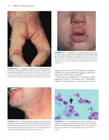
 mebooksfree.com  mebooksfree.com           mebooksfree.com          FIGURE 15–5    Staphylococcus aureus—Gram stain. Arrows               mebooksfree.com
                                                                                                            mebooksfree.com
                 FIGURE 15–3
                                Folliculitis. Note the multiple, small pustules on
                 the chin and neck. Staphylococcus aureus is the most common cause
                                                                     point to two “grapelike” clusters of gram-positive cocci. Arrowhead
                 of folliculitis. (Reproduced with permission from Wolff K, Goldsmith LA, Katz SI et
                                                                     points to neutrophil with pink segmented nuclei. (Used with permission
                                                                     from Professor Shirley Lowe, University of California, San Francisco School of
                 al (eds): Fitzpatrick’s Dermatology in General Medicine. 7th ed. New York: McGraw-Hill,
                 2008, pg 1699. Copyright © 2008 by The McGraw-Hill Companies, Inc.)
                                                                     Medicine.)


 mebooksfree.com  mebooksfree.com           mebooksfree.com                 mebooksfree.com                 mebooksfree.com                mebooksfree.com
   116   117   118   119   120   121   122   123   124   125   126