
Page 122 - Review of Medical Microbiology and Immunology ( PDFDrive )
P. 122
mebooksfree.com
mebooksfree.com
mebooksfree.com
mebooksfree.com
mebooksfree.com
mebooksfree.com
mebooksfree.com mebooksfree.com + mebooksfree.com Important Features 1 Typical Disease mebooksfree.com mebooksfree.com
mebooksfree.com
CHAPTER 15 Gram-Positive Cocci
111
TABLE 15–1 Staphylococci of Medical Importance
Species
Typical Hemolysis
Coagulase Production
S. aureus
β
Abscess, food poisoning, toxic shock syndrome
Protein A on surface
-
None
Sensitive to novobiocin
Infection of prosthetic heart valves and hips;
S. epidermidis
common member of skin flora
S. saprophyticus
1
All staphylococci are catalase-positive. - None Resistant to novobiocin Urinary tract
mebooksfree.com mebooksfree.com mebooksfree.com do not. Hemolysis of red cells by hemolysins produced by mebooksfree.com
mebooksfree.com
mebooksfree.com
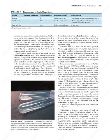
common and causes the most serious infections. Staphylo-
coccus aureus is distinguished from the others primarily by
S. aureus is the source of iron required for growth of the
coagulase production (Figure 15–6). Coagulase is an
organism. The iron in hemoglobin is recovered by the bacte-
enzyme that causes plasma to clot by activating prothrom-
ria and utilized in the synthesis of cytochrome enzymes used
to produce energy.
bin to form thrombin. Thrombin then catalyzes the activa-
tion of fibrinogen to form the fibrin clot. Staphylococcus
More than 90% of S. aureus strains contain plasmids
that encode a-lactamase, the enzyme that degrades many,
epidermidis and S. saprophyticus are often referred to as
coagulase-negative staphylococci.
tant to the β-lactamase–resistant penicillins, such as methi-
Staphylococcus aureus produces a carotenoid pigment
cillin and nafcillin, by virtue of changes in the
called staphyloxanthin, which imparts a golden color to its
penicillin-binding proteins (PBP) in their cell membrane.
colonies. This pigment enhances the pathogenicity of the but not all, penicillins. Some strains of S. aureus are resis-
mebooksfree.com
mebooksfree.com mebooksfree.com mebooksfree.com resistant S. aureus (MRSA) or nafcillin-resistant S. aureus mebooksfree.com
Genes on the bacterial chromosome called mecA genes
organism by inactivating the microbicidal effect of super-
mebooksfree.com
oxides and other reactive oxygen species within neutro-
encode these altered PBPs.
phils. Staphylococcus epidermidis does not synthesize this
These strains are commonly known as methicillin-
pigment and produces white colonies. The virulence of
(NRSA). MRSA causes both health care-acquired (HCA-
S. epidermidis is significantly less than that of S. aureus.
Two other characteristics further distinguish these species,
MRSA) and community-acquired (CA-MRSA) infections.
namely, S. aureus usually ferments mannitol and hemolyzes
MRSA currently accounts for more than 50% of S. aureus
strains isolated from hospital patients in the United States.
red blood cells, whereas S. epidermidis and S. saprophyticus
CA-MRSA is a very common cause of community-acquired
staphylococcal infections. Almost all strains of CA-MRSA
produce P-V leukocidin (see later) whereas relatively few
strains of HCA-MRSA do so. The most common strain of
MRSA in the United States is the “USA300” strain.
Strains of S. aureus with intermediate resistance to van-
mebooksfree.com mebooksfree.com mebooksfree.com encodes vancomycin resistance in S. aureus is the same as mebooksfree.com
mebooksfree.com
mebooksfree.com
comycin (VISA) and with full resistance to vancomycin
(VRSA) have also been detected. The cassette of genes that
the cassette that provides vancomycin resistance in entero-
cocci. These genes are located in a transposon on a plasmid
and encode the enzymes that substitute d-lactate for
d-alanine in the peptidoglycan.
S. aureus has several important cell wall components
and antigens:
(1) Protein A is the major protein in the cell wall. It is
an important virulence factor because it binds to the Fc
portion of IgG at the complement-binding site, thereby
mebooksfree.com
mebooksfree.com
mebooksfree.com mebooksfree.com mebooksfree.com A is used in certain tests in the clinical laboratory because mebooksfree.com
preventing the activation of complement. As a conse-
quence, no C3b is produced, and the opsonization and
phagocytosis of the organisms are greatly reduced. Protein
FIGURE 15–6
Coagulase test—Upper tube inoculated with
Staphylococcus aureus; lower tube inoculated with Staphylococcus
it binds to IgG and forms a “coagglutinate” with antigen–
epidermidis. Arrow points to clotted plasma formed by coagulase
antibody complexes. The coagulase-negative staphylococci
produced by S. aureus. (Used with permission from Professor Shirley Lowe, Uni-
do not produce protein A.
versity of California, San Francisco School of Medicine.)
mebooksfree.com mebooksfree.com mebooksfree.com mebooksfree.com mebooksfree.com mebooksfree.com