
Page 19 - Review of Medical Microbiology and Immunology ( PDFDrive )
P. 19
mebooksfree.com
mebooksfree.com
mebooksfree.com
mebooksfree.com
mebooksfree.com
mebooksfree.com mebooksfree.com mebooksfree.com The Gram stain is useful in two ways: mebooksfree.com mebooksfree.com
mebooksfree.com
mebooksfree.com
PART I Basic Bacteriology
8
GRAM STAIN
This staining procedure, developed in 1884 by the Danish
physician Christian Gram, is the most important procedure
(1) In the identification of many bacteria.
in microbiology. It separates most bacteria into two groups:
(2) In influencing the choice of antibiotic because, in gen-
the gram-positive bacteria, which stain blue, and the gram-
negative bacteria, which stain red. The Gram stain involves
G than are gram-negative bacteria.
the following four-step procedure: eral, gram-positive bacteria are more susceptible to penicillin
mebooksfree.com
mebooksfree.com mebooksfree.com mebooksfree.com be seen and describes the reason why. The alternative micro- mebooksfree.com
mebooksfree.com
However, not all bacteria can be seen in the Gram stain.
(1) The crystal violet dye stains all cells blue/purple.
Table 2–3 lists the medically important bacteria that cannot
(2) The iodine solution (a mordant) is added to form a
crystal violet–iodine complex; all cells continue to appear blue.
scopic approach to the Gram stain is also described.
(3) The organic solvent, such as acetone or ethanol,
Note that it takes approximately 100,000 bacteria/mL to see
extracts the blue dye complex from the lipid-rich, thin-
1 bacterium per microscopic field using the oil immersion
walled gram-negative bacteria to a greater degree than from
(100×) lens. So the sensitivity of the Gram stain procedure is
the lipid-poor, thick-walled gram-positive bacteria. The
low. This explains why a patient’s blood is rarely stained
gram-negative organisms appear colorless; the gram-positive
bacteria remain blue.
night to allow the bacteria to multiply. One important excep-
(4) The red dye safranin stains the decolorized gram-
tion to this is meningococcemia in which very high
negative cells red/pink; the gram-positive bacteria remain immediately but rather is incubated in blood cultures over-
mebooksfree.com mebooksfree.com mebooksfree.com Staphylococcus aureus, for example, five glycines link the mebooksfree.com
mebooksfree.com
mebooksfree.com
concentrations of Neisseria meningitidis can occur in the
blue.
blood.
make up the molecule. Synonyms for peptidoglycan are
murein and mucopeptide.
terminal D-alanine to the penultimate L-lysine.
Because peptidoglycan is present in bacteria but not in
Figure 2–5 illustrates the carbohydrate backbone, which
human cells, it is a good target for antibacterial drugs. Sev-
is composed of alternating N-acetylmuramic acid and
N-acetylglucosamine molecules. Attached to each of the
eral of these drugs, such as penicillins, cephalosporins, and
vancomycin, inhibit the synthesis of peptidoglycan by
muramic acid molecules is a tetrapeptide consisting of both
inhibiting the transpeptidase that makes the cross-links
D- and L-amino acids, the precise composition of which
differs from one bacterium to another. Two of these amino
between the two adjacent tetrapeptides (see Chapter 10).
acids are worthy of special mention: diaminopimelic acid,
Lysozyme, an enzyme present in human tears, mucus,
mebooksfree.com mebooksfree.com mebooksfree.com and saliva, can cleave the peptidoglycan backbone by mebooksfree.com
mebooksfree.com
mebooksfree.com
which is unique to bacterial cell walls, and d-alanine, which
is involved in the cross-links between the tetrapeptides and
breaking its glycosyl bonds, thereby contributing to the
in the action of penicillin. Note that this tetrapeptide con-
natural resistance of the host to microbial infection. Lyso-
zyme-treated bacteria may swell and rupture as a result of
tains the rare d-isomers of amino acids; most proteins
contain the l-isomer. The other important component in
the entry of water into the cells, which have a high internal
this network is the peptide cross-link between the two tet-
osmotic pressure. However, if the lysozyme-treated cells are
in a solution with the same osmotic pressure as that of the
rapeptides. The cross-links vary among species; in
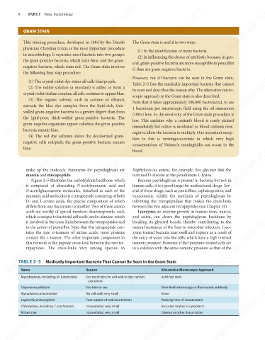
TABLE 2–3 Medically Important Bacteria That Cannot Be Seen in the Gram Stain
Name
Mycobacteria, including M. tuberculosis Reason Alternative Microscopic Approach
Acid-fast stain
Too much lipid in cell wall so dye cannot
penetrate
mebooksfree.com mebooksfree.com mebooksfree.com mebooksfree.com mebooksfree.com mebooksfree.com
Dark-field microscopy or fluorescent antibody
Too thin to see
Treponema pallidum
Mycoplasma pneumoniae
None
No cell wall; very small
Poor uptake of red counterstain
Legionella pneumophila
Prolong time of counterstain
Intracellular; very small
Chlamydiae, including C. trachomatis
Inclusion bodies in cytoplasm
Intracellular; very small
Giemsa or other tissue stains
Rickettsiae
mebooksfree.com mebooksfree.com mebooksfree.com mebooksfree.com mebooksfree.com mebooksfree.com