
Page 16 - Review of Medical Microbiology and Immunology ( PDFDrive )
P. 16
mebooksfree.com
mebooksfree.com
mebooksfree.com
mebooksfree.com
mebooksfree.com
mebooksfree.com mebooksfree.com electron mebooksfree.com mebooksfree.com of mebooksfree.com mebooksfree.com
mebooksfree.com
CHAPTER 2 Structure of Bacterial Cells 5
Red
Range of
Range of
Lower limit
optical
blood
microscope
microscope
human vision
cell
Escherichia coli
Mycoplasma
Bacillus
anthracis
Haemophilus
Hepatitis
influenzae
B virus
Candida Protozoa
Poxvirus
mebooksfree.com mebooksfree.com 0.03 0.05 0.1 0.3 0.5 Scale (µm) mebooksfree.com 100 300 mebooksfree.com
HIV
mebooksfree.com
albicans
mebooksfree.com
Poliovirus
10
5
30
3
1
0.005 0.01
50
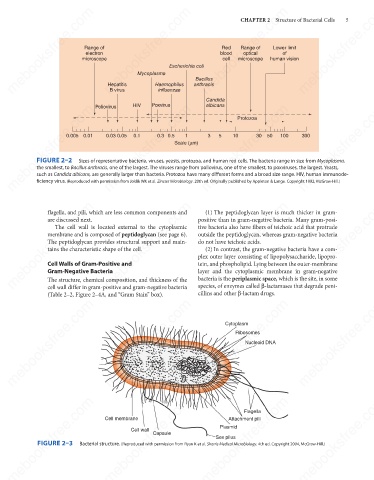
FIGURE 2–2
Sizes of representative bacteria, viruses, yeasts, protozoa, and human red cells. The bacteria range in size from Mycoplasma,
the smallest, to Bacillus anthracis, one of the largest. The viruses range from poliovirus, one of the smallest, to poxviruses, the largest. Yeasts,
such as Candida albicans, are generally larger than bacteria. Protozoa have many different forms and a broad size range. HIV, human immunode-
ficiency virus. (Reproduced with permission from Joklik WK et al. Zinsser Microbiology. 20th ed. Originally published by Appleton & Lange. Copyright 1992, McGraw-Hill.)
mebooksfree.com mebooksfree.com mebooksfree.com positive than in gram-negative bacteria. Many gram-posi- mebooksfree.com
mebooksfree.com
mebooksfree.com
flagella, and pili, which are less common components and
(1) The peptidoglycan layer is much thicker in gram-
are discussed next.
tive bacteria also have fibers of teichoic acid that protrude
The cell wall is located external to the cytoplasmic
outside the peptidoglycan, whereas gram-negative bacteria
membrane and is composed of peptidoglycan (see page 6).
The peptidoglycan provides structural support and main-
do not have teichoic acids.
tains the characteristic shape of the cell.
(2) In contrast, the gram-negative bacteria have a com-
plex outer layer consisting of lipopolysaccharide, lipopro-
Cell Walls of Gram-Positive and
tein, and phospholipid. Lying between the outer-membrane
Gram-Negative Bacteria
bacteria is the periplasmic space, which is the site, in some
The structure, chemical composition, and thickness of the
species, of enzymes called β-lactamases that degrade peni-
cell wall differ in gram-positive and gram-negative bacteria
cillins and other β-lactam drugs.
(Table 2–2, Figure 2–4A, and “Gram Stain” box). layer and the cytoplasmic membrane in gram-negative
mebooksfree.com mebooksfree.com mebooksfree.com mebooksfree.com mebooksfree.com mebooksfree.com
Cytoplasm
Ribosomes
Nucleoid DNA
mebooksfree.com
mebooksfree.com mebooksfree.com Cell membrane Capsule mebooksfree.com mebooksfree.com mebooksfree.com
Flagella
Attachment pili
Plasmid
Cell wall
Sex pilus
FIGURE 2–3
Bacterial structure. (Reproduced with permission from Ryan K et al. Sherris Medical Microbiology. 4th ed. Copyright 2004, McGraw-Hill.)
mebooksfree.com mebooksfree.com mebooksfree.com mebooksfree.com mebooksfree.com mebooksfree.com