
Page 197 - Review of Medical Microbiology and Immunology ( PDFDrive )
P. 197
mebooksfree.com
mebooksfree.com
mebooksfree.com
mebooksfree.com
mebooksfree.com
mebooksfree.com
mebooksfree.com
mebooksfree.com
mebooksfree.com mebooksfree.com mebooksfree.com in resistance and are not used for diagnostic purposes. mebooksfree.com
PART II Clinical Bacteriology
186
tuberculosis, or cause hematogenous dissemination result-
Circulating antibodies also form, but they play no role
ing in no immediate disease but with the risk of reactiva-
Patients deficient in cellular immunity, such as patients
tion in later life.
Figure 21–2 also describes secondary tuberculosis with
with acquired immunodeficiency syndrome (AIDS), are at
a cavity in the upper lobes. This can cause disease directly
much higher risk for disseminated, life-threatening tuber-
or result in reactivation disease in later life with central
another cause of defective cellular immunity that predis-
nervous system lesions, vertebral osteomyelitis (Pott’s dis-
ease), or involvement of other organs.
poses to severe tuberculosis. This emphasizes the impor-
Mycobacterium tuberculosis produces no exotoxins and culosis. Mutations in the interferon-γ receptor gene are
tance of activation of macrophages by interferon-γ in the
mebooksfree.com mebooksfree.com mebooksfree.com reaction. PPD is used as the antigen in the tuberculin skin mebooksfree.com
does not contain endotoxin in its cell wall. In fact, no
host defense against M. tuberculosis.
mebooksfree.com
mebooksfree.com
Prior infection can be detected by a positive tuberculin
mycobacteria produce toxins. The organism preferentially
skin test result, which is due to a delayed hypersensitivity
infects macrophages and other reticuloendothelial cells.
Mycobacterium tuberculosis survives and multiplies within
test. The intermediate-strength preparation of PPD, which
a cellular vacuole called a phagosome. The organism pro-
contains five tuberculin units, is usually used. The skin test
duces a protein called “exported repetitive protein” that
prevents the phagosome from fusing with the lysosome,
is evaluated by measuring the diameter of the induration
surrounding the skin test site (Figure 21–3). Note that
thereby allowing the organism to escape the degradative
induration (thickening), not simply erythema (reddening),
enzymes in the lysosome.
must be observed.
Lesions are dependent on the presence of the organism
and the host response. There are two types of lesions:
The diameter required to judge the test as positive varies
(1) Exudative lesions, which consist of an acute inflam-
Induration of 15 mm or more is positive in a person who
matory response and occur chiefly in the lungs at the initial depending on the status of the individual being tested.
has no known risk factors. Induration of 10 mm or more is
mebooksfree.com
mebooksfree.com
mebooksfree.com mebooksfree.com mebooksfree.com dents. Induration of 5 mm or more is positive in a person mebooksfree.com
site of infection.
positive in a person with high-risk factors, such as a home-
(2) Granulomatous lesions, which consist of a central
less person, intravenous drug users, or nursing home resi-
area of giant cells containing tubercle bacilli surrounded by
a zone of epithelioid cells. These giant cells, called Langhans’
who has deficient cell-mediated immunity (e.g., AIDS
giant cells, are an important pathologic finding in tuber-
patients) or has been in close contact with a person with
culous lesions. A tubercle is a granuloma surrounded by
active tuberculosis.
fibrous tissue that has undergone central caseation necro-
A positive skin test result indicates previous infection
sis. Tubercles heal by fibrosis and calcification.
by the organism but not necessarily active disease. The
The primary lesion of tuberculosis usually occurs in the
tuberculin test becomes positive 4 to 6 weeks after infection.
lungs. The parenchymal exudative lesion and the draining
lymph nodes together are called a Ghon complex. Primary
vaccine (see page 189) can cause a positive test, but the reac-
lesions usually occur in the lower lobes, whereas reactiva-
tions are usually only 5 to 10 mm and tend to decrease with
tion lesions usually occur in the apices. Reactivation lesions Immunization with bacillus Calmette-Guérin (BCG)
mebooksfree.com mebooksfree.com mebooksfree.com mebooksfree.com mebooksfree.com mebooksfree.com
also occur in other well-oxygenated sites such as the kid-
neys, brain, and bone. Reactivation is seen primarily in
immunocompromised or debilitated patients.
Spread of the organism within the body occurs by two
mechanisms:
(1) A tubercle can erode into a bronchus, empty its case-
ous contents, and thereby spread the organism to other
parts of the lungs, to the gastrointestinal tract if swallowed,
and to other persons if expectorated.
(2) It can disseminate via the bloodstream to many inter-
nal organs. Dissemination can occur at an early stage if cell-
mediated immunity fails to contain the initial infection or at
a late stage if a person becomes immunocompromised.
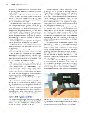
mebooksfree.com mebooksfree.com mebooksfree.com FIGURE 21–3 Tuberculin skin test. Purified protein derivative mebooksfree.com
mebooksfree.com
mebooksfree.com
Immunity & Hypersensitivity
After recovery from the primary infection, resistance to the
organism is mediated by cellular immunity (i.e., by CD4-
(PPD) was injected intradermally, and 48 hours later, the diameter of
positive T cells and macrophages). The CD4-positive T
induration was measured with a caliper. (Reproduced with permission from
cells are Th-1 helper T cells (see Chapter 58).
Talaro KP. Foundations in Microbiology. 8th ed. New York: McGraw-Hill, 2011.)
mebooksfree.com mebooksfree.com mebooksfree.com mebooksfree.com mebooksfree.com mebooksfree.com