
Page 430 - Review of Medical Microbiology and Immunology ( PDFDrive )
P. 430
mebooksfree.com
mebooksfree.com
mebooksfree.com
mebooksfree.com
mebooksfree.com mebooksfree.com mebooksfree.com mebooksfree.com mebooksfree.com mebooksfree.com
mebooksfree.com
mebooksfree.com
CHAPTER 50 Opportunistic Mycoses
419
B
A
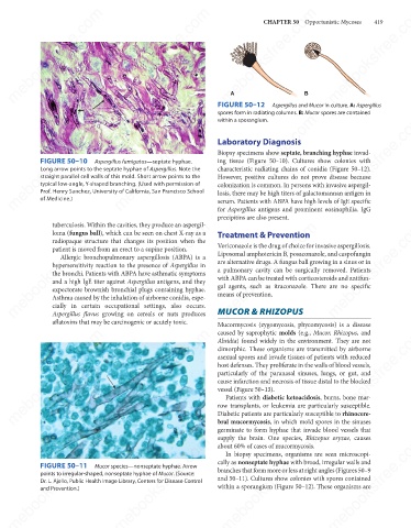
FIGURE 50–12
Aspergillus and Mucor in culture. A: Aspergillus
spores form in radiating columns. B: Mucor spores are contained
mebooksfree.com
mebooksfree.com mebooksfree.com Aspergillus fumigatus—septate hyphae. Laboratory Diagnosis mebooksfree.com mebooksfree.com
mebooksfree.com
within a sporangium.
Biopsy specimens show septate, branching hyphae invad-
FIGURE 50–10
ing tissue (Figure 50–10). Cultures show colonies with
characteristic radiating chains of conidia (Figure 50–12).
Long arrow points to the septate hyphae of Aspergillus. Note the
straight parallel cell walls of this mold. Short arrow points to the
However, positive cultures do not prove disease because
typical low-angle, Y-shaped branching. (Used with permission of
colonization is common. In persons with invasive aspergil-
Prof. Henry Sanchez, University of California, San Francisco School
losis, there may be high titers of galactomannan antigen in
of Medicine.)
serum. Patients with ABPA have high levels of IgE specific
for Aspergillus antigens and prominent eosinophilia. IgG
precipitins are also present.
mebooksfree.com
mebooksfree.com
mebooksfree.com mebooksfree.com mebooksfree.com Voriconazole is the drug of choice for invasive aspergillosis. mebooksfree.com
tuberculosis. Within the cavities, they produce an aspergil-
loma (fungus ball), which can be seen on chest X-ray as a
Treatment & Prevention
radiopaque structure that changes its position when the
patient is moved from an erect to a supine position.
Liposomal amphotericin B, posaconazole, and caspofungin
Allergic bronchopulmonary aspergillosis (ABPA) is a
are alternative drugs. A fungus ball growing in a sinus or in
hypersensitivity reaction to the presence of Aspergillus in
a pulmonary cavity can be surgically removed. Patients
the bronchi. Patients with ABPA have asthmatic symptoms
with ABPA can be treated with corticosteroids and antifun-
and a high IgE titer against Aspergillus antigens, and they
gal agents, such as itraconazole. There are no specific
expectorate brownish bronchial plugs containing hyphae.
means of prevention.
Asthma caused by the inhalation of airborne conidia, espe-
cially in certain occupational settings, also occurs.
Aspergillus flavus growing on cereals or nuts produces
aflatoxins that may be carcinogenic or acutely toxic. MUCOR & RHIZOPUS
Mucormycosis (zygomycosis, phycomycosis) is a disease
mebooksfree.com
mebooksfree.com
mebooksfree.com mebooksfree.com mebooksfree.com asexual spores and invade tissues of patients with reduced mebooksfree.com
caused by saprophytic molds (e.g., Mucor, Rhizopus, and
Absidia) found widely in the environment. They are not
dimorphic. These organisms are transmitted by airborne
host defenses. They proliferate in the walls of blood vessels,
particularly of the paranasal sinuses, lungs, or gut, and
cause infarction and necrosis of tissue distal to the blocked
vessel (Figure 50–13).
Patients with diabetic ketoacidosis, burns, bone mar-
row transplants, or leukemia are particularly susceptible.
Diabetic patients are particularly susceptible to rhinocere-
bral mucormycosis, in which mold spores in the sinuses
germinate to form hyphae that invade blood vessels that
supply the brain. One species, Rhizopus oryzae, causes
mebooksfree.com
mebooksfree.com
mebooksfree.com mebooksfree.com Mucor species—nonseptate hyphae. Arrow cally as nonseptate hyphae with broad, irregular walls and mebooksfree.com
mebooksfree.com
about 60% of cases of mucormycosis.
In biopsy specimens, organisms are seen microscopi-
FIGURE 50–11
branches that form more or less at right angles (Figures 50–9
points to irregular-shaped, nonseptate hyphae of Mucor. (Source:
and 50–11). Cultures show colonies with spores contained
Dr. L. Ajello, Public Health Image Library, Centers for Disease Control
within a sporangium (Figure 50–12). These organisms are
and Prevention.)
mebooksfree.com mebooksfree.com mebooksfree.com mebooksfree.com mebooksfree.com mebooksfree.com