
Page 559 - Review of Medical Microbiology and Immunology ( PDFDrive )
P. 559
mebooksfree.com
mebooksfree.com
mebooksfree.com
mebooksfree.com
mebooksfree.com mebooksfree.com Antigen Zone of equivalence antibody Antibody mebooksfree.com mebooksfree.com
mebooksfree.com
mebooksfree.com
mebooksfree.com
mebooksfree.com
PART VII Immunology
548
Zone of
Zone of
antigen
in well
excess
in well
excess
Agar plate
mebooksfree.com
mebooksfree.com mebooksfree.com mebooksfree.com antigen diffuses with time, precipitation rings form mebooksfree.com
mebooksfree.com
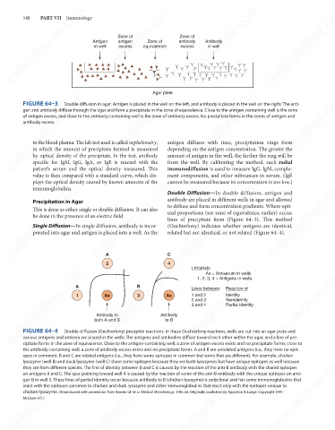
FIGURE 64–3
Double diffusion in agar. Antigen is placed in the well on the left, and antibody is placed in the well on the right. The anti-
gen and antibody diffuse through the agar and form a precipitate in the zone of equivalence. Close to the antigen-containing well is the zone
of antigen excess, and close to the antibody-containing well is the zone of antibody excess. No precipitate forms in the zones of antigen and
antibody excess.
in the blood plasma. The lab test used is called nephelometry,
depending on the antigen concentration. The greater the
in which the amount of precipitate formed is measured
by optical density of the precipitate. In the test, antibody
from the well. By calibrating the method, such radial
specific for IgM, IgG, IgA, or IgE is reacted with the
patient’s serum and the optical density measured. This
immunodiffusion is used to measure IgG, IgM, comple-
ment components, and other substances in serum. (IgE
value is then compared with a standard curve, which dis- amount of antigen in the well, the farther the ring will be
plays the optical density caused by known amounts of the
cannot be measured because its concentration is too low.)
mebooksfree.com
mebooksfree.com mebooksfree.com mebooksfree.com antibody are placed in different wells in agar and allowed mebooksfree.com
mebooksfree.com
immunoglobulins.
Double Diffusion—In double diffusion, antigen and
Precipitation in Agar
to diffuse and form concentration gradients. Where opti-
This is done as either single or double diffusion. It can also
mal proportions (see zone of equivalence, earlier) occur,
be done in the presence of an electric field.
lines of precipitate form (Figure 64–3). This method
Single Diffusion—In single diffusion, antibody is incor-
(Ouchterlony) indicates whether antigens are identical,
related but not identical, or not related (Figure 64–4).
porated into agar and antigen is placed into a well. As the
A
2 C LEGEND:
4
mebooksfree.com
mebooksfree.com mebooksfree.com 1 both A and B B Antibody mebooksfree.com mebooksfree.com mebooksfree.com
As = Antiserum in wells
1, 2, 3, 4 = Antigens in wells
A
Reaction of
Lines between
Identity
1 and 2
3
As
As
Nonidentity
2 and 3
Partial identity
3 and 4
Antibody to
to B
FIGURE 64–4
Double-diffusion (Ouchterlony) precipitin reactions. In these Ouchterlony reactions, wells are cut into an agar plate and
various antigens and antisera are placed in the wells. The antigens and antibodies diffuse toward each other within the agar, and a line of pre-
cipitate forms in the zone of equivalence. Close to the antigen-containing well, a zone of antigen excess exists and no precipitate forms; close to
the antibody-containing well, a zone of antibody excess exists and no precipitate forms. A and B are unrelated antigens (i.e., they have no epit-
opes in common). B and C are related antigens (i.e., they have some epitopes in common but some that are different). For example, chicken
mebooksfree.com mebooksfree.com mebooksfree.com mebooksfree.com mebooksfree.com mebooksfree.com
lysozyme (well B) and duck lysozyme (well C) share some epitopes because they are both lysozymes but have unique epitopes as well because
they are from different species. The line of identity between B and C is caused by the reaction of the anti-B antibody with the shared epitopes
on antigens B and C. The spur pointing toward well 4 is caused by the reaction of some of the anti-B antibody with the unique epitopes on anti-
gen B in well 3. These lines of partial identity occur because antibody to B (chicken lysozyme) is polyclonal and has some immunoglobulins that
react with the epitopes common to chicken and duck lysozyme and other immunoglobulins that react only with the epitopes unique to
chicken lysozyme. (Reproduced with permission from Brooks GF et al. Medical Microbiology. 19th ed. Originally published by Appleton & Lange. Copyright 1991
McGraw-Hill.)
mebooksfree.com mebooksfree.com mebooksfree.com mebooksfree.com mebooksfree.com mebooksfree.com