


Page 149 - Textbook of Pathology, 6th Edition
P. 149
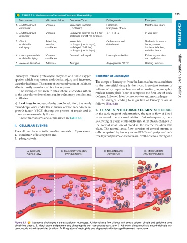
TABLE 6.1: Mechanisms of Increased Vascular Permeability. 133
Mechanism Microvasculature Response Type Pathogenesis Examples
1. Endothelial cell Venules Immediate transient Histamine, Mild thermal injury
contraction (15-30 min) bradykinin, others
2. Endothelial cell Venules Somewhat delayed (in 4-6 hrs) IL-1, TNF-α In vitro only CHAPTER 6
retraction prolonged (for 24 hrs or more)
3. Direct Arterioles, Immediate Cell necrosis and Moderate to severe
endothelial venules, prolonged (hrs to days), detachment burns, severe
cell injury capillaries or delayed (2-12 hrs) bacterial infection,
prolonged (hrs to days) radiation injury
4. Leucocyte-mediated Venules, Delayed, prolonged Leucocyte activation Pulmonary venules
endothelial injury capillaries and capillaries
5. Neovascularisation All levels Any type Angiogenesis, VEGF Healing, tumours
leucocytes release proteolytic enzymes and toxic oxygen Exudation of Leucocytes Inflammation and Healing
species which may cause endothelial injury and increased The escape of leucocytes from the lumen of microvasculature
vascular leakiness. This form of increased vascular leakiness to the interstitial tissue is the most important feature of
affects mostly venules and is a late response. inflammatory response. In acute inflammation, polymorpho-
The examples are seen in sites where leucocytes adhere
to the vascular endothelium e.g. in pulmonary venules and nuclear neutrophils (PMNs) comprise the first line of body
defense, followed later by monocytes and macrophages.
capillaries.
The changes leading to migration of leucocytes are as
v) Leakiness in neovascularisation. In addition, the newly follows (Fig. 6.4):
formed capillaries under the influence of vascular endothelial
growth factor (VEGF) during the process of repair and in 1. CHANGES IN THE FORMED ELEMENTS OF BLOOD.
tumours are excessively leaky. In the early stage of inflammation, the rate of flow of blood
These mechanisms are summarised in Table 6.1. is increased due to vasodilatation. But subsequently, there
is slowing or stasis of bloodstream. With stasis, changes in
II. CELLULAR EVENTS the normal axial flow of blood in the microcirculation take
place. The normal axial flow consists of central stream of
The cellular phase of inflammation consists of 2 processes: cells comprised by leucocytes and RBCs and peripheral cell-
1. exudation of leucocytes; and free layer of plasma close to vessel wall. Due to slowing and
2. phagocytosis.
Figure 6.4 Sequence of changes in the exudation of leucocytes. A, Normal axial flow of blood with central column of cells and peripheral zone
of cell-free plasma. B, Margination and pavementing of neutrophils with narrow plasmatic zone. C, Adhesion of neutrophils to endothelial cells with
pseudopods in the intercellular junctions. D, Emigration of neutrophils and diapedesis with damaged basement membrane.

