


Page 223 - Textbook of Pathology, 6th Edition
P. 223
Besides acute leukaemias,  other tumours in infancy and                                                  207
           childhood are: neuroblastoma, nephroblastoma (Wilms’
           tumour), retinoblastoma, hepatoblastoma, rhabdomyo-
           sarcoma, Ewing’s sarcoma, teratoma and CNS tumours.
           5. SEX.  Apart from the malignant tumours of organs
           peculiar to each sex, most tumours are generally more                                                      CHAPTER 8
           common in men than in women except cancer of the breast,
           gall bladder, thyroid and hypopharynx. Although there are
           geographic and racial variations, cancer of the breast is the
           commonest cancer in women throughout the world while lung
           cancer is the commonest cancer in men. The differences in
           incidence of certain cancers in the two sexes may be related
           to the presence of specific sex hormones.                                                                  Neoplasia

           B. Chronic Non-neoplastic (Pre-malignant)  Conditions
           Premalignant lesions are a group of conditions which
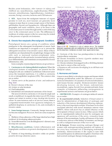
           predispose to the subsequent development of cancer. Such  Figure 8.16  Carcinoma  in situ of uterine cervix. The atypical
           conditions are important to recognise so as to prevent the  dysplastic squamous cells are confined to all the layers of the mucosa
           subsequent occurrence of an invasive cancer. Many of these  but the basement membrane on which these layers rest is intact.
           conditions are characterised by morphologic changes in the  ii) Cirrhosis of the liver has predisposition to develop
           cells such as increased nuclear-cytoplasmic ratio,  hepatocellular carcinoma.
           pleomorphism of cells and nuclei, increased mitotic activity,  iii) Chronic bronchitis in heavy cigarette smokers may
           poor differentiation, and sometimes accompanied by chronic  develop cancer of the bronchus.
           inflammatory cells.                                 iv) Chronic irritation from jagged tooth or ill-fitting denture
              Some examples of premalignant lesions are given below:
                                                               may lead to cancer of the oral cavity.
           1. Carcinoma in situ (intraepithelial neoplasia). When the  v) Squamous cell carcinoma developing in an old burn scar
           cytological features of malignancy are present but the  (Marjolin’s ulcer).
           malignant cells are confined to epithelium without invasion
           across the basement membrane, it is called as carcinoma  C. Hormones and Cancer
           in situ or intraepithelial neoplasia (CIN). The common sites  Cancer is more likely to develop in organs and tissues which
           are as under:                                       undergo proliferation under the influence of excessive
           i) Uterine cervix at the junction of ecto- and endocervix  hormonal stimulation. On cessation of hormonal stimulation,
           (Fig. 8.16)                                         such tissues become atrophic. Hormone-sensitive tissues
           ii) Bowen’s disease of the skin                     developing tumours are the breast, endometrium,
           iii) Actinic or solar keratosis                     myometrium, vagina, thyroid, liver, prostate and testis. Some
           iv) Oral leukoplakia                                examples of hormones influencing carcinogenesis in
           v) Intralobular and intraductal carcinoma of the breast.  experimental animals and humans are given below:
              The area involved in carcinoma in situ may be single and  1. OESTROGEN. Examples of oestrogen-induced cancers
           small, or multifocal. As regards the behaviour of CIN, it may  are as under:
           regress and return to normal or may develop into invasive  i) In experimental animals. Induction of breast cancer in
           cancer. In some instances such as in cervical cancer, there is  mice by administration of high-dose of oestrogen and
           a sequential transformation from squamous metaplasia, to  reduction of the tumour development following oophorec-
           epithelial dysplasia, to carcinoma in situ, and eventually to  tomy is the most important example. It has been known that
           invasive cancer.                                    associated infection with mouse mammary tumour virus
           2. Some benign tumours. Commonly, benign tumours do  (MMTV, Bittner milk factor) has an added influence on the
           not become malignant. However, there are some exceptions  development of breast cancer in mice. Other cancers which
           e.g.                                                can be experimentally induced in mice by oestrogens are
           i) Multiple villous adenomas of the large intestine have high  squamous cell carcinoma of the cervix, connective tissue
           incidence of developing adenocarcinoma.             tumour of the myometrium, Leydig cell tumour of the testis
                                                               in male mice, tumour of the kidney in hamsters, and benign
           ii) Neurofibromatosis (von Recklinghausen’s disease) may  as well as malignant tumours of the liver in rats.
           develop into sarcoma.
                                                               ii) In humans. Women receiving oestrogen therapy and
           3. Miscellaneous conditions. Certain inflammatory and  women with oestrogen-secreting granulosa cell tumour of
           hyperplastic conditions are prone to development of cancer,  the ovary have increased risk of developing endometrial
           e.g.                                                carcinoma. Adenocarcinoma of the vagina is seen with
           i) Patients of long-standing ulcerative colitis are predis-  increased frequency in adolescent daughters of mothers who
           posed to develop colorectal cancer.                 had received oestrogen therapy during pregnancy.

