


Page 219 - Textbook of Pathology, 6th Edition
P. 219
fragments or clusters of tumour cells break off to be carried                                            203
           in the coelomic fluid and are implanted elsewhere in the body
           cavity. Peritoneal cavity is involved most often, but
           occasionally pleural and pericardial cavities are also affected.
           A few examples of transcoelomic spread are as follows:
           a) Carcinoma of the stomach seeding to both ovaries                                                        CHAPTER 8
           (Krukenberg tumour).
           b) Carcinoma of the ovary spreading to the entire peritoneal
           cavity without infiltrating the underlying organs.
           c) Pseudomyxoma peritonei is the gelatinous coating of the
           peritoneum from mucin-secreting carcinoma of the ovary or
           apppendix.
           d) Carcinoma of the bronchus and breast seeding to the pleura                                              Neoplasia
           and pericardium.
           ii) Spread along epithelium-lined surfaces. It is unusual
           for a malignant tumour to spread along the epithelium-lined
           surfaces because intact epithelium and mucus coat are quite
           resistant to penetration by tumour cells. However,
           exceptionally a malignant tumour may spread through:
           a) the fallopian tube from the endometrium to the ovaries or
           vice-versa;
           b) through the bronchus into alveoli; and
           c) through the ureters from the kidneys into lower urinary
           tract.
           iii) Spread via cerebrospinal fluid. Malignant tumour of the
           ependyma and leptomeninges may spread by release of
           tumour fragments and tumour cells into the CSF and produce
           metastases at other sites in the central nervous system.
           iv) Implantation. Rarely, a tumour may spread by
           implantation by surgeon’s scalpel, needles, sutures, or may
           be implanted by direct contact such as transfer of cancer of
           the lower lip to the apposing upper lip.

           MECHANISM AND BIOLOGY OF
           INVASION AND METASTASIS

           The process of local invasion and distant spread by lymphatic
           and haematogenous routes discussed above involves passage
           through barriers before gaining access to the vascular lumen.
           This includes making the passage by the cancer cells by
           dissolution of extracellular matrix (ECM) at three levels— at
           the basement membrane of tumour itself, at the level of
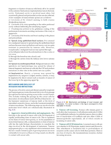
           interstitial connective tissue, and at the basement membrane  Figure 8.15  Mechanism and biology of local invasion and
                                                               metastasis.  The serial numbers in the figure correspond to their
           of microvasculature. The following steps are involved at the  description in the text.
           molecular level which are schematically illustrated in
           Fig. 8.15.                                          2. Tumour cell loosening. Normal cells remain glued to
                                                               each other due to presence of cell adhesion molecules (CAMs)
           1. Aggressive clonal proliferation and angiogenesis. The  i.e.E (epithelial)-cadherin. In epithelial cancers, there is either
           first step in the spread of cancer cells is the development of  loss or inactivation of E-cadherin and also other CAMs of
           rapidly proliferating clone of cancer cells. This is explained  immunoglobulin superfamily, all of which results in
           on the basis of tumour heterogeneity, i.e. in the population of  loosening of cancer cells.
           monoclonal tumour cells, a subpopulation or clone of tumour
           cells has the right biologic characteristics to complete the  3. Tumour cell-ECM interaction. Loosened cancer cells are
           steps involved in the development of metastasis. Tumour  now attached to ECM proteins, mainly laminin and fibronectin.
           angiogenesis plays a very significant role in metastasis since  This attachment is facilitated due to profoundness of
           the new vessels formed as part of growing tumour are more  receptors on the cancer cells for both these proteins. There is
           vulnerable to invasion as these evolving vessels are directly  also loss of integrins, the transmembrane receptors, further
           in contact with cancer cells.                       favouring invasion.

