


Page 226 - Textbook of Pathology, 6th Edition
P. 226
210


     SECTION I


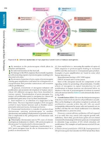
           Figure 8.19  Schematic representation of major properties of cancer in terms of molecular carcinogenesis.


              By mutation in the protooncogene which alters its  iii) Gene amplification i.e. increasing the number of copies of
           structure and function.                             DNA  sequence in protooncogene leading to increased
              By retroviral insertion in the host cell.        mDNA and thus increased or overexpressed gene product.
     General Pathology and Basic Techniques
              By damage to the DNA sequence that normally regulates  Examples of gene amplification are found in some solid
           growth-promoting signals of protooncogenes resulting in its  human tumours e.g.
           abnormal activation.                                   Neuroblastoma having n-MYC HSR region.
              By erroneous formation of extra copies of protooncogene  ERB-B1 in breast and ovarian cancer.
           causing gene amplification and hence its overexpression or  Most of the oncogenes encode for components of cell
           overproduction that promotes autonomous and excessive  signaling system for promoting cell proliferation. Possible
           cellular proliferation.                             effects of oncogenes in signal transduction for cell
               In general, overactivity of oncogenes enhances cell  proliferation in human tumours are discussed below in
           proliferation and promotes development of human cancer.  relation to the role of protooncogenes in mitosis in normal
           About 100 different oncogenes have been described in  cell cycle and are listed in Table 8.4 and schematically shown
           various cancers. Transformation of proto-oncogene (i.e.  in Fig. 8.20:
           normal cell proliferation gene) to oncogenes (i.e. cancer cell
           proliferation gene) may occur by three mechanisms:  i) Growth factors (GFs). GFs were the first protoonocgenes
           i) Point mutations i.e. an alteration of a single base in the  to be discovered which encode for cell proliferation cascade.
           DNA chain. The most important example is RAS oncogene  They act by binding to cell surface receptors to activate cell
           carried in many human tumours such as bladder cancer,  proliferation cascade within the cell. GFs are small
           pancreatic adenocarcinoma, cholangiocarcinoma.      polypeptides elaborated by many cells and normally act on
           ii) Chromosomal translocations i.e. transfer of a portion of one  another cell than the one which synthesised it to stimulate
           chromosome carrying protooncogene to another        its proliferation i.e. paracrine action.
           chromosome and making it independent of growth controls.  However, a cancer cell may synthesise a GF and respond
           This is implicated in the pathogenesis of leukaemias and  to it as well; this way cancer cells acquire growth self-
           lymphomas e.g.                                      sufficiency. Most often, growth factor genes are not altered
              Philadelphia chromosome seen in 95% cases of chronic  or mutated but instead growth factor genes are overexpressed
           myelogenous leukaemia in which c-ABL protooncogene on  to stimulate large secretion of GFs which stimulate cell
           chromosome 9 is translocated to chromosome 22.      proliferation. The examples of such tumour secreted GFs are
              In 75% cases of Burkitt’s lymphoma, translocation of c-  as under:
           MYC proto-oncogene from its site on chromosome 8 to a  a) Platelet-derived growth factor- (PDGF-β): Overexpression of
           portion on chromosome 14.                           SIS protooncogene that encodes for PDGF-β and thus there

