


Page 229 - Textbook of Pathology, 6th Edition
P. 229
213
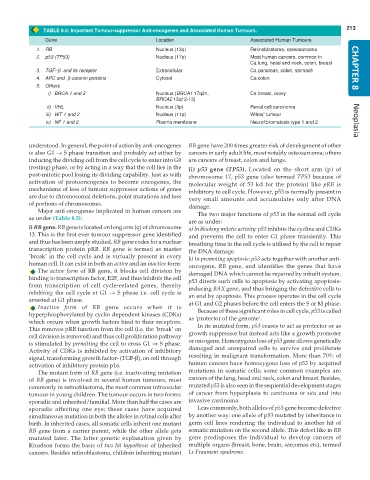
TABLE 8.5: Important Tumour-suppressor Anti-oncogenes and Associated Human Tumours.
Gene Location Associated Human Tumours
1. RB Nucleus (13q) Retinoblastoma, osteosarcoma
2. p53 (TP53) Nucleus (17p) Most human cancers, common in
Ca lung, head and neck, colon, breast
3. TGF–β and its receptor Extracellular Ca pancreas, colon, stomach CHAPTER 8
4. APC and β-catenin proteins Cytosol Ca colon
5. Others
i) BRCA 1 and 2 Nucleus (BRCA1 17q21, Ca breast, ovary
BRCA2 13q12-13)
ii) VHL Nucleus (3p) Renal cell carcinoma
iii) WT 1 and 2 Nucleus (11p) Wilms’ tumour
iv) NF 1 and 2 Plasma membrane Neurofibromatosis type 1 and 2 Neoplasia
understood. In general, the point of action by anti-oncogenes RB gene have 200 times greater risk of development of other
is also G1 → S phase transition and probably act either by cancers in early adult life, most notably osteosarcoma; others
inducing the dividing cell from the cell cycle to enter into G0 are cancers of breast, colon and lungs.
(resting) phase, or by acting in a way that the cell lies in the ii) p53 gene (TP53). Located on the short arm (p) of
post-mitotic pool losing its dividing capability. Just as with chromosome 17, p53 gene (also termed TP53 because of
activation of protooncogenes to become oncogenes, the molecular weight of 53 kd for the protein) like pRB is
mechanisms of loss of tumour suppressor actions of genes inhibitory to cell cycle. However, p53 is normally present in
are due to chromosomal deletions, point mutations and loss very small amounts and accumulates only after DNA
of portions of chromosomes. damage.
Major anti-oncogenes implicated in human cancers are The two major functions of p53 in the normal cell cycle
as under (Table 8.5):
are as under:
i) RB gene. RB gene is located on long arm (q) of chromosome a) In blocking mitotic activity: p53 inhibits the cyclins and CDKs
13. This is the first ever tumour suppressor gene identified and prevents the cell to enter G1 phase transiently. This
and thus has been amply studied. RB gene codes for a nuclear breathing time in the cell cycle is utilised by the cell to repair
transcription protein pRB. RB gene is termed as master the DNA damage.
‘break’ in the cell cycle and is virtually present in every b) In promoting apoptosis: p53 acts together with another anti-
human cell. It can exist in both an active and an inactive form: oncogene, RB gene, and identifies the genes that have
The active form of RB gene, it blocks cell division by damaged DNA which cannot be repaired by inbuilt system.
binding to transcription factor, E2F, and thus inhibits the cell p53 directs such cells to apoptosis by activating apoptosis-
from transcription of cell cycle-related genes, thereby inducing BAX gene, and thus bringing the defective cells to
inhibiting the cell cycle at G1 → S phase i.e. cell cycle is an end by apoptosis. This process operates in the cell cycle
arrested at G1 phase. at G1 and G2 phases before the cell enters the S or M phase.
Inactive form of RB gene occurs when it is
Because of these significant roles in cell cycle, p53 is called
hyperphosphorylated by cyclin dependent kinases (CDKs) as ‘protector of the genome’.
which occurs when growth factors bind to their receptors.
This removes pRB function from the cell (i.e. the ‘break’ on In its mutated form, p53 ceases to act as protector or as
cell division is removed) and thus cell proliferation pathway growth suppressor but instead acts like a growth promoter
is stimulated by permitting the cell to cross G1 → S phase. or oncogene. Homozygous loss of p53 gene allows genetically
Activity of CDKs is inhibited by activation of inhibitory damaged and unrepaired cells to survive and proliferate
signal, transforming growth factor- (TGF-β), on cell through resulting in malignant transformation. More than 70% of
activation of inhibitory protein p16. human cancers have homozygous loss of p53 by acquired
The mutant form of RB gene (i.e. inactivating mutation mutations in somatic cells; some common examples are
of RB gene) is involved in several human tumours, most cancers of the lung, head and neck, colon and breast. Besides,
commonly in retinoblastoma, the most common intraocular mutated p53 is also seen in the sequential development stages
tumour in young children. The tumour occurs in two forms: of cancer from hyperplasia to carcinoma in situ and into
sporadic and inherited/familial. More than half the cases are invasive carcinoma.
sporadic affecting one eye; these cases have acquired Less commonly, both alleles of p53 gene become defective
simultaneous mutation in both the alleles in retinal cells after by another way: one allele of p53 mutated by inheritance in
birth. In inherited cases, all somatic cells inherit one mutant germ cell lines rendering the individual to another hit of
RB gene from a carrier parent, while the other allele gets somatic mutation on the second allele. This defect like in RB
mutated later. The latter genetic explanation given by gene predisposes the individual to develop cancers of
Knudson forms the basis of two hit hypothesis of inherited multiple organs (breast, bone, brain, sarcomas etc), termed
cancers. Besides retinoblastoma, children inheriting mutant Li-Fraumeni syndrome.

