


Page 230 - Textbook of Pathology, 6th Edition
P. 230
214 iii) Transforming growth factor-ββ ββ β (TGF-ββ ββ β) and its receptor. inherited as a mutation in VHL tumour suppressor gene
Normally, TGF-β is significant inhibitor of cell proliferation, located on chromosome 3p. This results in activation of genes
especially in epithelial, endothelial and haematopoieitc cells that promote angiogenesis, survival and proliferation; VHL
It acts by binding to TGF-β receptor and then the complex so gene is found inactivated in 60% cases of renal cell carcinoma.
formed acts in G1 phase of cell cycle at two levels: c) Wilms’ tumour (WT) gene: Both WT1 an WT2 genes are
It activates CDK inhibitors (CDKIs) with growth located on chromosome 11 and normally prevent neoplastic
inhibitory effect. proliferation of cells in embryonic kidney. Mutant form of
It suppresses the growth prmoter genes such as MYC, WT-1 and 2 are seen in hereditary Wilms’ tumour.
CDKs and cyclins. d) Neurofibroma (NF) gene: NF genes normally prevent
SECTION I
Mutant form of TGF-β gene or its receptor impairs the proliferation of Schwann cells. Two mutant forms are
growth inhibiting effect and thus permits cell proliferation. described: NF1 and NF2 seen in neurofibromatosis type 1
Examples of mutated form of TGF-β are seen in cancers of and type 2.
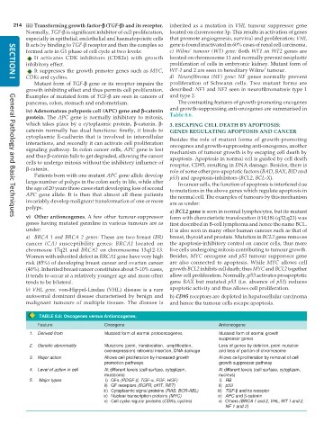
pancreas, colon, stomach and endometrium. The contrasting features of growth-promoting oncogenes
iv) Adenomatous polyposis coli (APC) gene and ββ ββ β-catenin and growth-suppressing anti-oncogenes are summarised in
protein. The APC gene is normally inhibitory to mitosis, Table 8.6.
which takes place by a cytoplasmic protein, β-catenin. β- 3. ESCAPING CELL DEATH BY APOPTOSIS:
catenin normally has dual functions: firstly, it binds to GENES REGULATING APOPTOSIS AND CANCER
cytoplasmic E-cadherin that is involved in intercellular Besides the role of mutant forms of growth-promoting
interactions, and secondly it can activate cell proliferation oncogenes and growth-suppressing anti-oncogenes, another
signaling pathway. In colon cancer cells, APC gene is lost mechanism of tumour growth is by escaping cell death by
and thus β-catenin fails to get degraded, allowing the cancer apoptosis. Apoptosis in normal cell is guided by cell death
cells to undergo mitosis without the inhibitory influence of receptor, CD95, resulting in DNA damage. Besides, there is
β-catenin. role of some other pro-apoptotic factors (BAD, BAX, BID and
Patients born with one mutant APC gene allele develop p53) and apoptosis-inhibitors (BCL2, BCL-X).
large number of polyps in the colon early in life, while after In cancer cells, the function of apoptosis is interfered due
the age of 20 years these cases start developing loss of second to mutations in the above genes which regulate apoptosis in
APC gene allele. It is then that almost all these patients the normal cell. The examples of tumours by this mechanism
invariably develop malignant transformation of one or more are as under:
polyps.
a) BCL2 gene is seen in normal lymphocytes, but its mutant
v) Other antioncogenes. A few other tumour-suppressor form with characteristic translocation (t14;18) (q32;q21) was
General Pathology and Basic Techniques
genes having mutated germline in various tumours are as first described in B-cell lymphoma and hence the name BCL.
under: It is also seen in many other human cancers such as that of
a) BRCA 1 and BRCA 2 genes: These are two breast (BR) breast, thyroid and prostate. Mutation in BCL2 gene removes
cancer (CA) susceptibility genes: BRCA1 located on the apoptosis-inhibitory control on cancer cells, thus more
chromosoe 17q21 and BRCA2 on chromosome 13q12-13. live cells undergoing mitosis contributing to tumour growth.
Women with inherited defect in BRCA1 gene have very high Besides, MYC oncogene and p53 tumour suppressor gene
risk (85%) of developing breast cancer and ovarian cancer are also connected to apoptosis. While MYC allows cell
(40%). Inherited breast cancer constitutes about 5-10% cases, growth BCL2 inhibits cell death; thus MYC and BCL2 together
it tends to occur at a relatively younger age and more often allow cell proliferation. Normally, p53 activates proapoptotic
tends to be bilateral. gene BAX but mutated p53 (i.e. absence of p53) reduces
b) VHL gene. von-Hippel-Lindau (VHL) disease is a rare apoptotic activity and thus allows cell proliferation.
autosomal dominant disease characterised by benign and b) CD95 receptors are depleted in hepatocellular carcinoma
malignant tumours of multiple tissues. The disease is and hence the tumour cells escape apoptosis.
TABLE 8.6: Oncogenes versus Antioncogenes.
Feature Oncogene Antioncogene
1. Derived from Mutated form of normal protooncogenes Mutated form of normal growth
suppressor genes
2. Genetic abnormality Mutations (point, translocation, amplification, Loss of genes by deletion, point mutation
overexpression) retroviral insertion, DNA damage and loss of portion of chromosome
3. Major action Allows cell proliferation by increased growth Allows cell proliferation by removal of cell
promotion pathways growth suppressor pathway
4. Level of action in cell At different levels (cell surface, cytoplasm, At different levels (cell surface, cytoplasm,
mutations) nucleus)
5. Major types i) GFs (PDGF-β, TGF-α, FGF, HGF) i) RB
ii) GF receptors (EGFR, cKIT, RET) ii) p53
iii) Cytoplasmic signal proteins (RAS, BCR-ABL) iii) TGF-β and its receptor
iv) Nuclear transcription proteins (MYC) iv) APC and β-catenin
v) Cell cycle regular proteins (CDKs, cyclins) v) Others (BRCA 1 and 2, VHL, WT 1 and 2,
NF 1 and 2)

