


Page 249 - Textbook of Pathology, 6th Edition
P. 249
fine needle aspiration cytology (FNAC). The superficial masses fixed paraffin sections or cytological smears. The complex 233
can be aspirated under direct vision while deep-seated of antigen-antibody on slide is made visible for light
masses such as intra-abdominal, pelvic organs and microscopic identification by either fluorescent dyes (‘fluoro-
retroperitoneum are frequently investigated by ultrasound chromes’) or by enzyme system (‘chromogens’). The specific
(US) or computed tomography (CT)-guided fine needle antibody against a particular cellular antigen is obtained by
aspirations. The smears are fixed in 95% ethanol by wet hybridoma technique for monoclonal antibody production.
fixation, or may be air-dried unfixed. While Papanicolaou These monoclonal antibodies, besides being specific against CHAPTER 8
method of staining is routinely employed in most laboratories antigen, are highly sensitive in detection of antigenic
for wet fixed smears, others prefer H and E due to similarity component, and, therefore, impart objectivity to the
in staining characteristics in the sections obtained by paraffin- subjective tumour diagnosis made by the surgical
embedding. Air-dried smears are stained by May-Grunwald- pathologist.
Giemsa or Leishman stain. FNAC has a diagnostic reliability Though the list of immunohistochemical stains is ever
between 80-97% but it must not be substituted for clinical increasing, an important group of antibody stains directed
judgement or compete with an indicated histopathologic against various classes of intermediate filaments is useful in Neoplasia
biopsy. classification of poorly-differentiated tumours of epithelial
or mesenchymal origin (Table 8.13). This subject is discussed
3. Histochemistry and Cytochemistry already in Chapter 2 and an abbreviated list of antibody stains
in some common cancers of unknown origin is given in
Histochemistry and cytochemistry are additional diagnostic Table 2.3.
tools which help the pathologist in identifying the chemical
composition of cells, their constituents and their products 5. Electron Microscopy
by special staining methods.
Though immunohistochemical techniques are more Ultrastructural examination of tumour cells offers selective
useful for tumour diagnosis (see below), histochemical and role in diagnostic pathology. EM examination may be helpful
cytochemical methods are still employed for this purpose. in confirming or substantiating a tumour diagnosis arrived
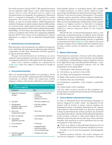
Some of the common examples are summarised in at by light microscopy and immunohistochemistry. A few
Table 8.12, while the subject is discussed at length in general features of malignant tumour cells by EM
Chapter 2. examination can be appreciated:
i) Cell junctions, their presence and type.
4. Immunohistochemistry ii) Cell surface, e.g. presence of microvilli.
This is an immunological method of recognising a cell by iii) Cell shape and cytoplasmic extensions.
one or more of its specific components in the cell membrane, iv) Shape of the nucleus and features of nuclear membrane.
cytoplasm or nucleus. These cell components (called v) Nucleoli, their size and density.
antigens) combine with specific antibodies on the formalin-
vi) Cytoplasmic organelles—their number is generally
reduced.
TABLE 8.12: Common Histochemical/Cytochemical Stains
in Tumour Diagnosis. vii) Dense bodies in the cytoplasm.
viii) Any other secretory product in the cytoplasm e.g.
Substance Stain
melanosomes in melanoma and membrane-bound granules
1. Basement membrane/ • Periodic acid-Schiff (PAS) in endocrine tumours.
collagen • Reticulin
• Van Gieson 6. Tumour Markers (Biochemical Assays)
• Masson’s trichrome
In order to distinguish from the preceding techniques of
2. Glycogen • PAS with diastase loss
tumour diagnosis in which ‘stains’ are imparted on the
3. Glycoproteins, • PAS with diastase tumour cells in section or smear, tumour markers are
glycolipids, glycomucins persistence biochemical assays of products elaborated by the tumour
(epithelial origin)
cells in blood or other body fluids. It is, therefore, pertinent
4. Acid mucin • Alcian blue to keep in mind that many of these products are produced
(mesenchymal origin)
by normal body cells too, and thus the biochemical estimation
5. Mucin (in general) • Combined Alcian blue-PAS of the product in blood or other fluid reflects the total
6. Argyrophilic/ • Silver stains substance and not by the tumour cells alone. These methods,
argentaffin granules therefore, lack sensitivity as well as specificity and can only
be employed for the following:
7. Cross striations • PTAH stain
Firstly, as an adjunct to the pathologic diagnosis arrived
8. Enzymes • Myeloperoxidase at by other methods and not for primary diagnosis of cancer.
• Acid phosphatase
• Alkaline phosphatase Secondly, it can be used for prognostic and therapeutic
purposes.
9. Nucleolar organiser • Colloidal silver stain
regions (NORs) Tumour markers include: cell surface antigens (or
oncofoetal antigens), cytoplasmic proteins, enzymes,

