


Page 273 - Textbook of Pathology, 6th Edition
P. 273
and Herpes simplex) during pregnancy is associated with                                                  257
           multisystem anomalies and TORCH syndrome in the
           newborn (page 191).
           6. Congenital syphilis. As discussed in Chapter 6, vertical
           transmission of syphilis from mother to foetus is
           characterised by Hutchinson’s triad: interstitial keratitis,                                               CHAPTER 10
           sensorineural deafness and deformed Hutchinson’s teeth,
           along with saddle-nose deformity.

                   CYTOGENETIC (KARYOTYPIC)
                          ABNORMALITIES

           Human germ cells (ova and sperms) contain 23 chromo-
           somes (haploid or 1N) while all the nucleated somatic cells
           of the human body contain 23 pairs of chromosomes
           (diploid or 2N)—44 autosomes and 2 sex chromosomes,
           being XX in females (46, XX) and XY in males (46, XY). The
           branch of science dealing with the study of human                                                          Genetic and Paediatric Diseases
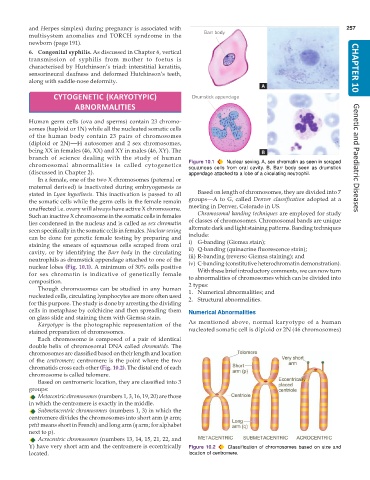
           chromosomal abnormalities is called cytogenetics    Figure 10.1  Nuclear sexing. A, sex chromatin as seen in scraped
                                                               squamous cells from oral cavity. B, Barr body seen as drumstick
           (discussed in Chapter 2).                           appendage attached to a lobe of a circulating neutrophil.
              In a female, one of the two X chromosomes (paternal or
           maternal derived) is inactivated during embryogenesis as
           stated in Lyon hypothesis. This inactivation is passed to all  Based on length of chromosomes, they are divided into 7
           the somatic cells while the germ cells in the female remain  groups—A to G, called Denver  classification adopted at a
           unaffected i.e. ovary will always have active X chromosome.  meeting in Denver, Colorado in US.
           Such an inactive X chromosome in the somatic cells in females  Chromosomal banding techniques are employed for study
           lies condensed in the nucleus and is called as sex chromatin  of classes of chromosomes. Chromosomal bands are unique
                                                               alternate dark and light staining patterns. Banding techniques
           seen specifically in the somatic cells in females. Nuclear sexing
           can be done for genetic female testing by preparing and  include:
           staining the smears of squamous cells scraped from oral  i) G-banding (Giemsa stain);
           cavity, or by identifying the Barr body in the circulating  ii) Q-banding (quinacrine fluorescence stain);
           neutrophils as drumstick appendage attached to one of the  iii) R-banding (reverse Giemsa staining); and
           nuclear lobes (Fig. 10.1). A minimum of 30% cells positive  iv) C-banding (constitutive heterochromatin demonstration).
           for sex chromatin is indicative of genetically female  With these brief introductory comments, we can now turn
           composition.                                        to abnormalities of chromosomes which can be divided into
                                                               2 types:
              Though chromosomes can be studied in any human   1. Numerical abnormalities; and
           nucleated cells, circulating lymphocytes are more often used  2. Structural abnormalities.
           for this purpose. The study is done by arresting the dividing
           cells in metaphase by colchicine and then spreading them  Numerical Abnormalities
           on glass slide and staining them with Giemsa stain.
              Karyotype is the photographic representation of the  As mentioned above, normal karyotype of a human
           stained preparation of chromosomes.                 nucleated somatic cell is diploid or 2N (46 chromosomes)
              Each chromosome is composed of a pair of identical
           double helix of chromosomal DNA called chromatids. The
           chromosomes are classified based on their length and location
           of the  centromere; centromere is the point where the two
           chromatids cross each other (Fig. 10.2). The distal end of each
           chromosome is called telomere.
              Based on centromeric location, they are classified into 3
           groups:
              Metacentric chromosomes (numbers 1, 3, 16, 19, 20) are those
           in which the centromere is exactly in the middle.
              Submetacentric chromosomes (numbers 1, 3) in which the
           centromere divides the chromosomes into short arm (p arm;
           petit means short in French) and long arm (q arm; for alphabet
           next to p).
              Acrocentric chromosomes (numbers 13, 14, 15, 21, 22, and
           Y) have very short arm and the centromere is eccentrically  Figure 10.2  Classification of chromosomes based on size and
           located.                                            location of centromere.

